. 2009 Nov;83(22):11966-78.
doi: 10.1128/JVI.01515-09. Epub 2009 Sep 9.

Antagonism to and intracellular sequestration of human tetherin by the human immunodeficiency virus type 2 envelope glycoprotein

Affiliations

Antagonism to and intracellular sequestration of human tetherin by the human immunodeficiency virus type 2 envelope glycoprotein

Anna Le Tortorec et al. J Virol. 2009 Nov.

Abstract

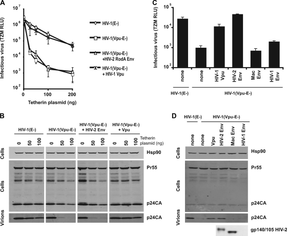
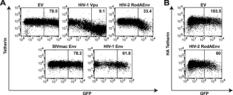
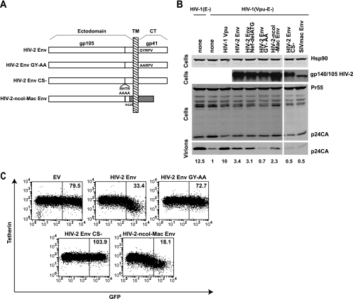
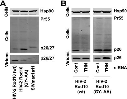
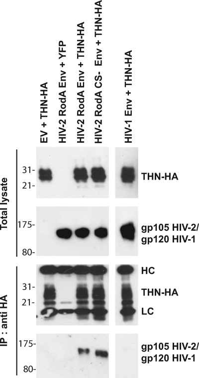
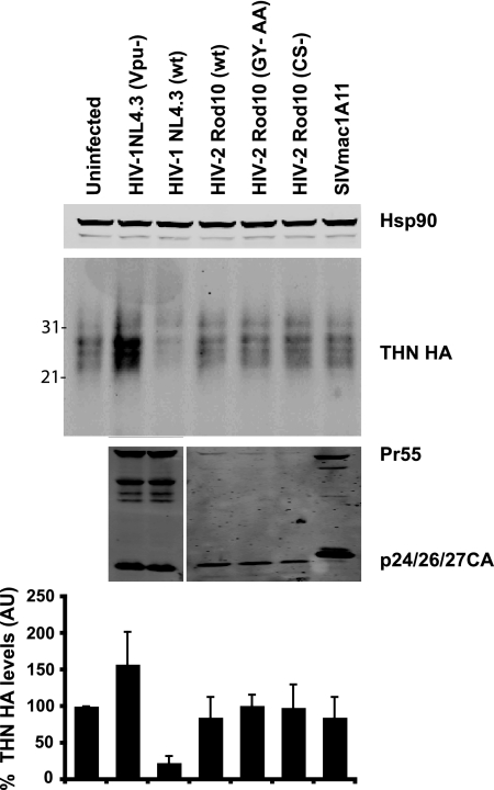
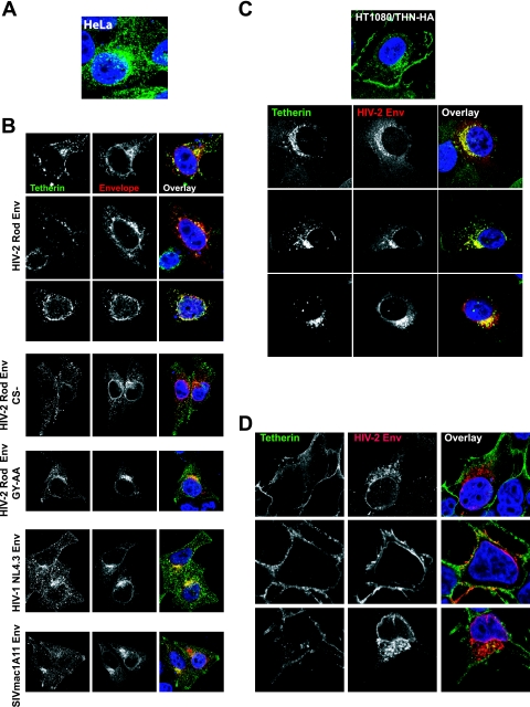
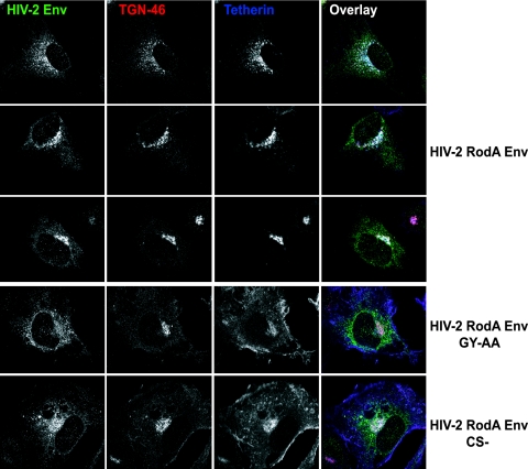
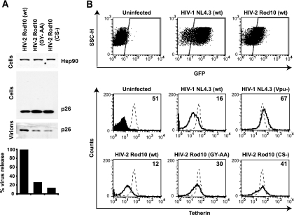
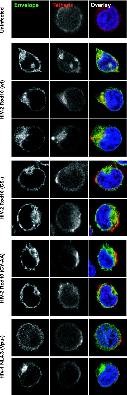
Tetherin (CD317/BST-2), an interferon-induced membrane protein, restricts the release of nascent retroviral particles from infected cell surfaces. While human immunodeficiency virus type 1 (HIV-1) encodes the accessory gene vpu to overcome the action of tetherin, the lineage of primate lentiviruses that gave rise to HIV-2 does not. It has been previously reported that the HIV-2 envelope glycoprotein has a Vpu-like function in promoting virus release. Here we demonstrate that the HIV-2 Rod envelope glycoprotein (HIV-2 Rod Env) is a tetherin antagonist. Expression of HIV-2 Rod Env, but not that of HIV-1 or the closely related simian immunodeficiency virus (SIV) SIVmac1A11, counteracts tetherin-mediated restriction of Vpu-defective HIV-1 in a cell-type-specific manner. This correlates with the ability of the HIV-2 Rod Env to mediate cell surface downregulation of tetherin. Antagonism requires an endocytic motif conserved across HIV/SIV lineages in the gp41 cytoplasmic tail, but specificity for tetherin is governed by extracellular determinants in the mature Env protein. Coimmunoprecipitation studies suggest an interaction between HIV-2 Rod Env and tetherin, but unlike studies with Vpu, we found no evidence of tetherin degradation. In the presence of HIV-2 Rod Env, tetherin localization is restricted to the trans-Golgi network, suggesting Env-mediated effects on tetherin trafficking sequester it from virus assembly sites on the plasma membrane. Finally, we recapitulated these observations in HIV-2-infected CD4+ T-cell lines, demonstrating that tetherin antagonism and sequestration occur at physiological levels of Env expression during virus replication.


Figures

FIG. 1.
FIG. 1.
Cell-type-dependent antagonism of tetherin restriction by the HIV-2 RodA envelope. (A) 293T cells were transfected with envelope-defective HIV-1 (E-) or a Vpu-defective counterpart, HIV-1 (Vpu-E-) molecular clones, in the presence of HIV-2 RodA Env or HIV-1 Vpu expression vectors and increasing doses of a human tetherin expression vector and pseudotyped with VSV-G. Supernatants were harvested 48 h later and used to infect HeLa-TZM indicator cells. Infectious virus release is plotted as β-galactosidase activity in relative light units (RLU) based on a commercial chemiluminescence assay. Error bars represent standard deviations of the means of three independent experiments. (B) Cell lysates of 293T cells in panel A and virions pelleted from the corresponding supernatants were subjected to SDS-PAGE and Western blotting for HIV-1 Gag or cellular Hsp90 and revealed by Licor fluorescent secondary antibodies. (C) A similar experiment as that in panel A was performed in HeLa cells, which express tetherin endogenously. In this case expression constructs for the HIV-1 NL4.3 Env and SIVmac1A11 Env were also used. Error bars represent standard deviations of the means of three independent experiments. (D) Western blots corresponding to results in panel C for HIV-1 p24-CA and HIV-2 Env gp140/105 (in cell lysates).
FIG. 2.
FIG. 2.
HIV-2 RodA Env expression mediates cell surface tetherin downregulation in HeLa cells. HeLa (A) or 293T/THN-HA (B) cells were transfected with a control empty vector (EV) or vectors encoding the indicated viral proteins linked to enhanced GFP via an IRES. At 48 h posttransfection the cells were stained for cell surface tetherin levels with a monoclonal anti-BST-2 antibody (A) or a monoclonal anti-HA (B) antibody and then a secondary goat-anti-mouse Alexa 633 antibody. The cells were then analyzed by flow cytometry. Median fluorescence intensities are indicated for the boxed regions.
FIG. 3.
FIG. 3.
Determinants of tetherin antagonism in HIV-2 Env. (A) HIV-2 RodA Env constructs were made with inactivating mutations in the GYxxθ motif (GY 719/720 AA): RodA GY-AA, disruption of the SU-TM cleavage site RTHR, RodA-CS-, and a chimeric envelope in which the RodA and SIVmac1A11 Envs were fused at a conserved NcoI site in the extracellular gp41 coding sequence (Rod-NcoI-Mac). (B) The ability of these Envs to promote Vpu virus release from HeLa cells was tested as described for Fig. 1, and Western blot assays were performed on cell lysates and virions for HIV-1 p24-CA and HIV-2 Env. Numbers represent the fold increase in virus release (p24 band intensity) compared to the Vpu-defective control. (C) HeLa cells were transfected with Env mutant and chimera IRES-GFP constructs, and cell surface staining for tetherin was analyzed by flow cytometry and compared to that for the control empty vector (EV) or wt RodA Env (reproduced from Fig. 2). Median fluorescence intensities are indicated for the boxed regions.
FIG. 4.
FIG. 4.
Effects of Env on tetherin-mediated particle release of HIV-2 Rod10 virions. (A) HeLa cells were infected with VSV-G-pseudotyped HIV-2 Rod10, HIV-2 Rod10 (GY-AA), or SIVmac1A11 at an MOI of 0.2 as standardized on HeLa-TZM. Forty-eight hours later virions and cell lysates were analyzed by Western blotting for HIV-2/SIVmac p26/p27 CA. (B) HeLa cells were treated with siRNA oligonucleotides directed at human tetherin or a control pool and then infected with VSV-G-pseudotyped HIV-2 Rod10 and HIV-2 Rod10 (GY-AA) and processed as described for panel A.
FIG. 5.
FIG. 5.
Coimmunoprecipitation of HIV-2 RodA Env with tetherin. HeLa cells were transfected with the indicated plasmid vectors with empty vector (EV) or yellow fluorescent protein (YFP) vector replacing Env or tetherin-HA, respectively, as negative controls. Forty-eight hours later, tetherin was immunoprecipitated (IP) from cell lysates and subjected to SDS-PAGE. Total lysates and immunoprecipitates were then Western blotted for tetherin-HA and HIV-2 or HIV-1 Env. HC and LC signify the heavy and light chains of the immunoprecipitating antibody, respectively.
FIG. 6.
FIG. 6.
Tetherin is not degraded in HIV-2-infected cells. HT1080 cells stably expressing tetherin-HA (HT/THN-HA) were infected with VSV-G-pseudotyped HIV-1 (wt), HIV-1 (Vpu-), HIV-2 (wt), HIV-2 (GY-AA), HIV-2 (CS-), or SIVmac1A11 at an MOI of 2 as standardized on HeLa-TZM cells to ensure approximately 90% of the cells were infected. At 48 h after infection total cell lysates were subjected to SDS-PAGE and Western blotted for tetherin-HA, HIV-1/HIV-2 CA, and Hsp90. Corresponding tetherin band intensities were corrected for Hsp90 levels and plotted as a percentage of tetherin-HA levels relative to the uninfected cells. Error bars represent standard deviations of three independent experiments. AU, arbitrary units.
FIG. 7.
FIG. 7.
Localization of tetherin in HeLa, HT1080, and 293T cells in response to envelope expression. (A) Localization of tetherin in HeLa cells stained with a mouse polyclonal anti-human BST-2 antibody (green). (B) Effects of envelope expression on endogenous tetherin localization in HeLa cells. Cells were transfected with the indicated envelope and 24 h later fixed and stained for tetherin (green) and envelope (red). (C) Tetherin-HA localization in HT1080/THN-HA cells expressing HIV-2 RodA Env introduced by retroviral vector transduction. Tetherin was stained with anti-HA (green) and envelope was costained (red). (D) 293T-THN/HA cells transfected with HIV-2 RodA Env were stained as described above. Nuclei were counterstained with DAPI (blue).
FIG. 8.
FIG. 8.
HIV-2 Env induces tetherin sequestration in the trans-Golgi network. HT1080/THN-HA cells expressing HIV-2 RodA Env or the indicated mutant were stained for HIV-2 Env (green), the TGN marker TGN46 (red), and tetherin-HA (blue).
FIG. 9.
FIG. 9.
Antagonism and cell surface downregulation of tetherin by HIV-2 in CD4+ T-cell lines. (A) Jurkat cells were infected with the indicated HIV-2 Rod10 virus pseudotyped with VSV-G at an MOI of 0.2. Cell lysates and pellet virions were analyzed by Western blotting 48 h later, and virus release was quantified as the supernatant p26-CA band intensity, as a percentage of the wild-type control. (B) CEM-G37 cells were infected with the indicated VSV-G-pseudotyped viral stock at an MOI of 1. Forty-eight hours later cells were stained for surface tetherin expression and analyzed by flow cytometry. GFP+ infected cells were gated, and surface tetherin levels (solid lines) were compared to those of uninfected CEM-G37 cells (dotted lines). Numbers indicate median fluorescence intensities of surface tetherin on the infected cells. The solid peak in the upper left histogram represents the binding of the isotype control.
FIG. 10.
FIG. 10.
Intracellular sequestration of tetherin in HIV-2-infected Jurkat cells. Jurkat cells were infected with the indicated VSV-G-pseudotyped viral stocks at an MOI of 1. Forty-eight hours later the cells were allowed to adhere to polylysine-coated coverslips, fixed, permeabilized, and stained for HIV-2/HIV-1 Env (green) or tetherin (red) using Alexa 488 and 633 secondary antibodies, respectively. Nuclei were counterstained with DAPI (blue), and the cells were examined by confocal microscopy.

References

    1. Abada, P., B. Noble, and P. M. Cannon. 2005. Functional domains within the human immunodeficiency virus type 2 envelope protein required to enhance virus production. J. Virol. 79:3627-3638. - PMC - PubMed
    1. Bartee, E., A. McCormack, and K. Fruh. 2006. Quantitative membrane proteomics reveals new cellular targets of viral immune modulators. PLoS Pathog. 2:e107. - PMC - PubMed
    1. Boge, M., S. Wyss, J. S. Bonifacino, and M. Thali. 1998. A membrane-proximal tyrosine-based signal mediates internalization of the HIV-1 envelope glycoprotein via interaction with the AP-2 clathrin adaptor. J. Biol. Chem. 273:15773-15778. - PubMed
    1. Bour, S., H. Akari, E. Miyagi, and K. Strebel. 2003. Naturally occurring amino acid substitutions in the HIV-2 ROD envelope glycoprotein regulate its ability to augment viral particle release. Virology 309:85-98. - PubMed
    1. Bour, S., U. Schubert, K. Peden, and K. Strebel. 1996. The envelope glycoprotein of human immunodeficiency virus type 2 enhances viral particle release: a Vpu-like factor? J. Virol. 70:820-829. - PMC - PubMed

Publication types

LinkOut - more resources