. 2009 Nov;83(22):11502-13.
doi: 10.1128/JVI.01318-09. Epub 2009 Sep 9.

Histone deacetylases 1 and 2 are phosphorylated at novel sites during varicella-zoster virus infection

Affiliations

Histone deacetylases 1 and 2 are phosphorylated at novel sites during varicella-zoster virus infection

Matthew S Walters et al. J Virol. 2009 Nov.

Abstract

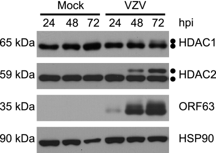
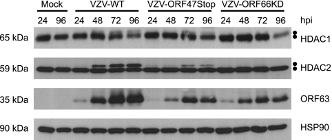
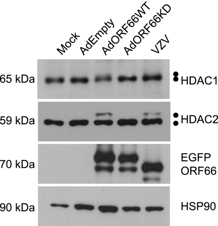
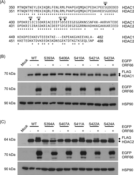
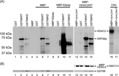
ORF66p, a virion-associated varicella-zoster virus (VZV) protein, is a member of a conserved Alphaherpesvirinae kinase family with homology to herpes simplex virus US3 kinase. Expression of ORF66p in cells infected with VZV or an adenovirus expressing only ORF66p results in hyperphosphorylation of histone deacetylase 1 (HDAC1) and HDAC2. Mapping studies reveal that phosphorylation is at a unique conserved Ser residue in the C terminus of both HDACs. This modification requires an active kinase domain in ORF66p, as neither protein is phosphorylated in cells infected with VZV lacking kinase activity. However, hyperphosphorylation appears to occur indirectly, as within the context of in vitro kinase reactions, purified ORF66p phosphorylates a peptide derived from ORF62p, a known substrate, but does not phosphorylate HDAC. These results support a model where ORF66p is necessary but not sufficient to effect hyperphosphorylation of HDAC1 and HDAC2.


Figures

FIG. 1.
FIG. 1.
Kinetic analysis of HDAC1 and HDAC2 forms during VZV infection. MeWo cells were mock infected or infected with cell-associated wild-type VZV at a multiplicity of infection of 0.025 and harvested 24, 48, and 72 hpi. At 72 hpi, all of the cells were infected as determined by phase microscopy and/or ORF66p-GFP expression. Equal amounts of total protein (20 μg) from each sample were analyzed by Western blotting using antisera specific for HDAC1, HDAC2, ORF63, and HSP90.
FIG. 2.
FIG. 2.
Analysis of HDAC1 and HDAC2 in infected-cell extracts following incubation with CIAP. MeWo cells were mock infected or infected with cell-associated wild-type VZV at a multiplicity of infection of 0.025 and harvested at 72 hpi. At 72 hpi, all of the cells were infected as determined by phase microscopy and/or ORF66p-GFP expression. Lysates from mock- and VZV-infected cells were reacted with dephosphorylation buffer alone (untreated), CIAP, or inactivated CIAP as described in Materials and Methods. Equal amounts of total protein (20 μg) from each sample were analyzed by Western blotting using antisera specific for HDAC1, HDAC2, and HSP90.
FIG. 3.
FIG. 3.
Analysis of HDAC1 and HDAC2 hyperphosphorylation in cells infected with wild-type and kinase-deficient VZV. MeWo cells were mock infected or infected with cell-associated wild-type (VZV-WT), ORF47p kinase-deficient (VZV-ORF47Stop), or ORF66p kinase-deficient (VZV-ORF66KD) VZV at a multiplicity of infection of 0.025 and harvested 24, 48, 72, and 96 hpi. At 96 hpi, all of the cells were infected as determined by phase microscopy and/or ORF66p-GFP expression. Equal amounts of total protein (20 μg) from each sample were analyzed by Western blotting using antisera specific for HDAC1, HDAC2, ORF63, and HSP90.
FIG. 4.
FIG. 4.
Analysis of HDAC1 and HDAC2 in cells infected with adenoviruses expressing wild-type and kinase-deficient ORF66p. MeWo cells were mock infected or infected with AdEmpty, AdORF66WT, or AdORF66KD at a multiplicity of infection of 10 in conjunction with AdTetOff at a multiplicity of infection of 5. As a positive control, MeWo cells were infected with cell-associated wild-type VZV at a multiplicity of infection of 0.025. At 48 hpi, cells were harvested and equal amounts of total protein (20 μg) were analyzed by Western blotting using antisera specific for HDAC1, HDAC2, GFP, and HSP90.
FIG. 5.
FIG. 5.
Identification of Ser residues in HDAC1 and HDAC2 that are phosphorylated in response to ORF66p expression. (A) Amino acid sequence alignment of the C termini of HDAC1 and HDAC2. The amino acid sequences of HDAC1 and HDAC2 are aligned, and positions marked with asterisks identify identical amino acids. Inverted triangles indicate phosphorylation sites in both proteins. The numbers above each triangle correspond to residues in HDAC1. (B) To map the ORF66p target phosphorylation site(s) in HDAC1, 293A cells were mock transformed or cotransformed with 1 μg of either pFLAG-HDAC1, pFLAG-S393A, pFLAG-S406A, pFLAG-S410A, pFLAG-S421A, or pFLAG-S423A and 1 μg of pEGFP-C1 or pEGFP-ORF66. After 48 h, cells were harvested and equal amounts of total protein (20 μg) from each sample were analyzed by Western blotting using antisera specific for FLAG, GFP, and HSP90. (C) To map the ORF66p target phosphorylation site(s) in HDAC2, 293A cells were mock transformed or cotransformed with 1 μg of pFLAG-HDAC2, pFLAG-S394A, pFLAG-S407A, pFLAG-S411A, pFLAG-S422A, or pFLAG-S424A and 1 μg of pEGFP or pEGFP-ORF66. After 48 h, cells were harvested and equal amounts of total protein (20 μg) from each sample were analyzed by Western blotting using antisera specific for FLAG, GFP, and HSP90.
FIG. 6.
FIG. 6.
In vitro analysis of ORF66p-dependent HDAC hyperphosphorylation. (A) Purified MBP substrate and GST kinase proteins were incubated in optimal kinase buffer, subsequently separated on 7% polyacrylamide denaturing gels, and then processed for autoradiography as described in Materials and Methods. Lanes 1 to 4 contain GST, GST66WT, GST66KD, and MBP incubated alone, respectively. Lanes 5 to 7 contain MBP incubated with GST, GST66WT, and GST66KD, respectively. The bracket in lane 10 identifies phosphorylated MBP-62pep. Lane 8 contains MBP-62pep incubated alone. Lanes 9 to 11 contain MBP-62pep incubated with GST, GST66WT, and GST66KD, respectively. Lane 12 contains MBP-HDAC2WT incubated alone. Lanes 13 to 15 contain MBP-HDAC2WT incubated with GST, GST66WT, and GST66KD, respectively. Lanes 16 and 17 contain CKII incubated with MBP-HDAC2WT and MBP-HDAC2MUT, respectively. (B) Equivalent levels of GST and MBP were detected in lanes 1 to 15 by Western blot analysis of the membranes following autoradiography. For lanes 16 and 17, Coomassie brilliant blue staining of the gel prior to drying and autoradiography was used to demonstrate equivalent levels of MBPs in each reaction mixture.
FIG. 7.
FIG. 7.
Plaquing efficiency and plaque size of ORF66p kinase-deficient VZV in cells treated with sodium butyrate. (A) MeWo cells were mock treated or pretreated with 1 mM sodium butyrate for 6 h. Following treatment, cells were infected with serial dilutions of cell-free wild-type (VZV-WT) or ORF66p kinase-deficient (VZV-ORF66KD) VZV. Cells were maintained in medium in the absence or presence of 1 mM sodium butyrate. After 6 days, monolayers were fixed and stained and plaques were counted. Relative plaquing efficiency was calculated as described in the text. Each column represents the average from two independent experiments, and error bars indicate standard deviations. A star indicates a P value [t(pval) = 0.0003] as determined by t test. (B) MeWo cells on coverslips were pretreated and infected as described above. Medium was replaced and fresh drug was added daily. Three days postinfection, cells were fixed, stained for gE, and observed by immunofluorescence microscopy. For each virus sample, images of 20 plaques were taken with a 10× objective and the size of each plaque was quantified using ImageJ software. The relative plaque size for each virus was calculated as described in the text. Results from a representative experiment are shown. Error bars indicate standard deviations. A star indicates a P value [t(pval) = 0.00008] as determined by t test. (C) Representative images for the average relative plaque size for each virus in the absence (Mock) and presence of 1 mM sodium butyrate. Images were captured at 3 days postinfection using a 10× objective following fixation and staining for gE by immunofluorescence microscopy.
FIG. 8.
FIG. 8.
Effect of inhibition of HDAC activity on growth and viral gene expression in cells infected with ORF66p kinase-deficient VZV. (A) MeWo cells were mock treated or pretreated with 1 mM sodium butyrate for 6 h. Following treatment, cells were infected with cell-free wild-type (VZV-WT) or ORF66p kinase-deficient (VZV-ORF66KD) VZV at a multiplicity of infection of 0.0001. Cells were maintained in medium with or without 1 mM sodium butyrate as indicated, and medium was replaced daily. At 24-h intervals postinfection, cell-associated virus titers were measured by titration on fresh monolayers of MeWo cells. Five days postinfection, monolayers were fixed and stained and plaques were counted to calculate the titers of infectious centers. Each datum point represents the average from two independent experiments, and the error bars indicate standard deviations. Stars identify P values [t(pval) < 0.006] for VZV-ORF66KD at 72, 96, and 120 hpi, as determined by t test. (B) MeWo cells were pretreated as described previously and infected with cell-free wild-type (VZV-WT) or ORF66p kinase-deficient (VZV-ORF66KD) VZV at a multiplicity of infection of 0.0001. Postinfection, cells were maintained as described above. At the indicated time points postinfection, cells were harvested and equal amounts of total protein (20 μg) were analyzed by Western blotting using antisera specific for ORF4p, ORF61p, ORF62p, ORF63p, and actin.

Similar articles

Cited by

References

    1. Arvin, A. M. 1996. Varicella-zoster virus. Clin. Microbiol. Rev. 9:361-381. - PMC - PubMed
    1. Benetti, L., and B. Roizman. 2004. Herpes simplex virus protein kinase US3 activates and functionally overlaps protein kinase A to block apoptosis. Proc. Natl. Acad. Sci. USA 101:9411-9416. - PMC - PubMed
    1. Besser, J., M. Ikoma, K. Fabel, M. H. Sommer, L. Zerboni, C. Grose, and A. M. Arvin. 2004. Differential requirement for cell fusion and virion formation in the pathogenesis of varicella-zoster virus infection in skin and T cells. J. Virol. 78:13293-13305. - PMC - PubMed
    1. Bradford, M. M. 1976. A rapid and sensitive method for the quantitation of microgram quantities of protein utilizing the principle of protein-dye binding. Anal. Biochem. 72:248-254. - PubMed
    1. Carpenter, J. E., E. P. Henderson, and C. Grose. 2009. Enumeration of an extremely high particle-to-PFU ratio for varicella-zoster virus. J. Virol. 83:6917-6921. - PMC - PubMed

Publication types