. 2009 Nov;83(22):11726-33.
doi: 10.1128/JVI.01463-09. Epub 2009 Sep 9.

Tryptophan residues in the portal protein of herpes simplex virus 1 critical to the interaction with scaffold proteins and incorporation of the portal into capsids

Affiliations

Tryptophan residues in the portal protein of herpes simplex virus 1 critical to the interaction with scaffold proteins and incorporation of the portal into capsids

Kui Yang et al. J Virol. 2009 Nov.

Abstract

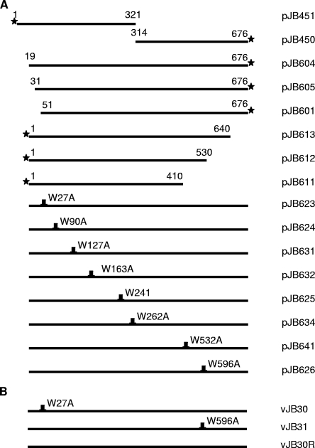
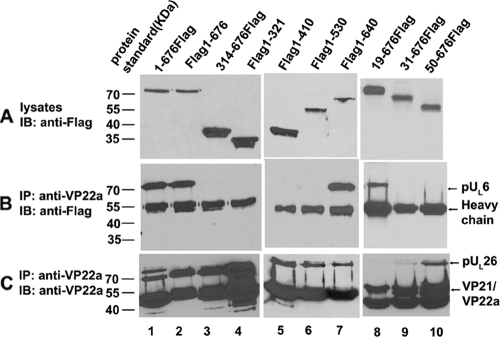
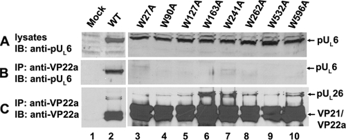
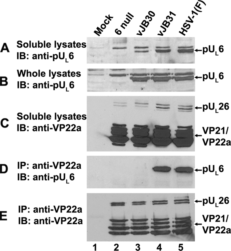
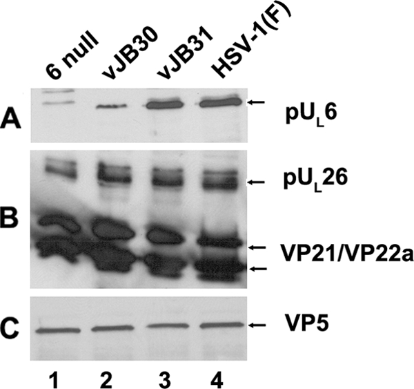
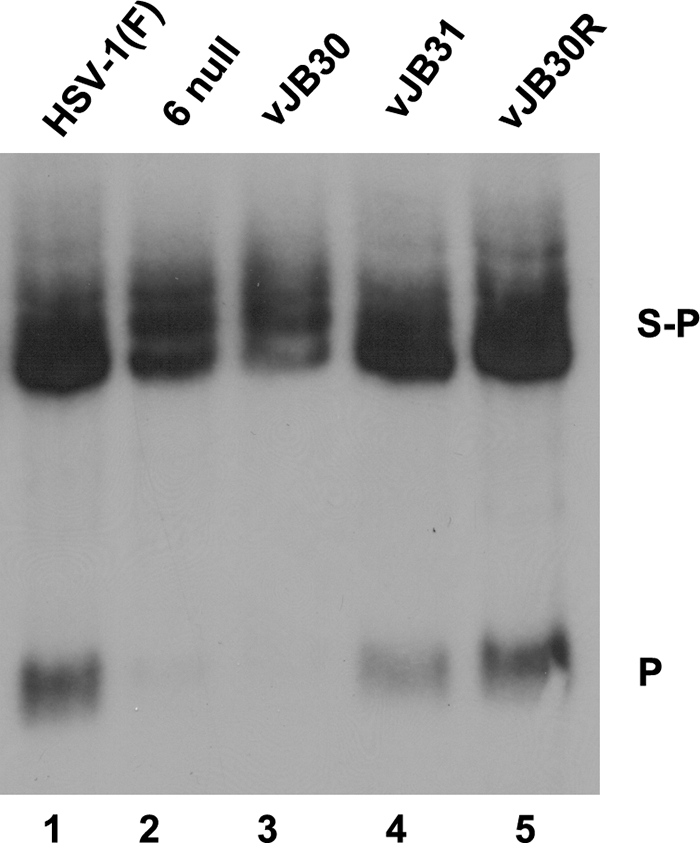
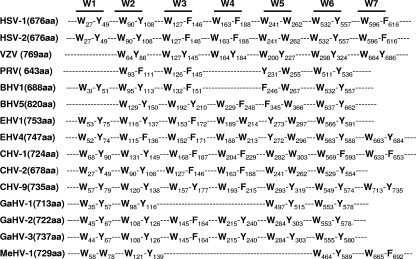
Incorporation of the herpes simplex virus 1 (HSV-1) portal vertex into the capsid requires interaction with a 12-amino-acid hydrophobic domain within capsid scaffold proteins. The goal of this work was to identify domains and residues in the UL6-encoded portal protein pUL6 critical to the interaction with scaffold proteins. We show that whereas the wild-type portal and scaffold proteins readily coimmunoprecipitated with one another in the absence of other viral proteins, truncation beyond the first 18 or last 36 amino acids of the portal protein precluded this coimmunoprecipitation. The coimmunoprecipitation was also precluded by mutation of conserved tryptophan (W) residues to alanine (A) at positions 27, 90, 127, 163, 241, 262, 532, and 596 of UL6. All of these W-to-A mutations precluded the rescue of a viral deletion mutant lacking UL6, except W163A, which supported replication poorly, and W596A, which fully rescued replication. A recombinant virus bearing the W596A mutation replicated and packaged DNA normally, and scaffold proteins readily coimmunoprecipitated with portal protein from lysates of infected cells. Thus, viral functions compensated for the W596A mutation's detrimental effects on the portal-scaffold interaction seen during transient expression of portal and scaffold proteins. In contrast, the W27A mutation precluded portal-scaffold interactions in infected cell lysates, reduced the solubility of pUL6, decreased incorporation of the portal into capsids, and abrogated viral-DNA cleavage and packaging.


Figures

FIG. 1.
FIG. 1.
Diagrams of pUL6 expression plasmid (A) and recombinant virus (B) with mutations in the UL6 gene. The designation of the plasmid (A) or recombinant virus (B) is indicated to the right of each line. The number above each line is the codon number. Letters plus numbers above each line indicate single-amino-acid substitutions. The stars designate the positions of the Flag epitope tags.
FIG. 2.
FIG. 2.
Mapping regions of pUL6 required for scaffold protein interactions. CV1 cells were cotransfected with expression plasmids encoding full-length pUL26 and Flag-tagged pUL6 or its derivatives. (A) Twenty-four hours after transfection, cells were harvested and lysed in RIPA buffer. The lysates were electrophoretically separated, transferred to nitrocellulose, and probed with anti-Flag antibody to monitor pUL6-Flag expression. (B) The lysates were reacted with anti-scaffold protein monoclonal antibody. The presence of pUL6 in immunoprecipitated material was detected by immunoblotting. (C) Immunoprecipitation reactions were performed with anti-scaffold antibody followed by immunoblotting with the same antibody. In all cases, bound immunoglobulins were revealed by enhanced chemiluminescence. The positions of pUL6, scaffold proteins, and the mouse immunoglobulin G heavy chain are indicated on the right. The positions of protein mass standards are indicated on the left. IP, immunoprecipitation; IB, immunoblotting.
FIG. 3.
FIG. 3.
Replacement of tryptophan by alanine within pUL6 abolishes its interaction with scaffold protein in transfected cells. CV1 cells were cotransfected with expression plasmids bearing full-length UL26 and wild-type UL6 or UL6 genes containing substitutions of alanine for tryptophan codons. Twenty-four hours after transfection, cells were harvested and lysed in RIPA buffer, and coimmunoprecipitation with anti-ICP35 monoclonal antibodies was performed. (A) Immunoblots of transfected cell lysates probed with pUL6-specific antibody. (B) Immunoblot of proteins immunoprecipitated with anti-scaffold antibody and probed with pUL6-specific antibody. (C) Immunoblot of proteins immunoprecipitated with anti-scaffold antibody and probed with the same antibody. The positions of pUL6 and scaffold proteins are indicated on the right. Bound immunoglobulins were revealed by enhanced chemiluminescence. IP, immunoprecipitation; IB, immunoblotting.
FIG. 4.
FIG. 4.
pUL6 and scaffold protein interaction in virus-infected cells. CV1 cells were mock infected or infected with HSV-1(F), vJB30(W27A), vJB31(W596A), or a UL6 null mutant. Eighteen hours after infection, coimmunoprecipitation was performed using anti-scaffold antibodies. (A) Immunoblot of electrophoretically separated lysates probed with anti-pUL6 antibody. (B) Whole-cell lysates probed with anti-pUL6 antibody. (C) Soluble lysates probed with anti-scaffold antibody. (D) Immunoblot of material immunoprecipitated with anti-scaffold antibody and probed with antibody to pUL6. (E) Immunoblot of proteins immunoprecipitated with scaffold-specific antibody and probed with the same antibody. Bound immunoglobulins were revealed by enhanced chemiluminescence. The positions of scaffold proteins and pUL6 are indicated on the right. IP, immunoprecipitation; IB, immunoblotting.
FIG. 5.
FIG. 5.
B-capsid proteins probed with pUL6-, VP22a-, or VP5-specific antibodies. CV1 cells (4 × 10⁸) were infected with 5 PFU/cell of HSV-1(F), UL6 null, vJB30(W27A), or vJB31(W596A). Twenty hours after infection, B capsids were purified, denatured, separated by SDS-12% polyacrylamide gel electrophoresis, and transferred onto a nitrocellulose membrane, followed by immunoblotting with anti-pUL6, anti-VP22a, or anti-VP5 antibodies. Bound immunoglobulin was revealed by enhanced chemiluminescence.
FIG. 6.
FIG. 6.
Southern blotting of viral genomes digested with BamHI. CV1 cells (2 × 10⁶) were infected with 5 PFU/cell of HSV-1(F), UL6 null, JB30(W27A), vJB31(W596A), or vJB30R. Fifteen hours after infection, total nuclear DNA was extracted, digested with BamHI, transferred to a nylon membrane (0.45 μM), and hybridized with radiolabeled BamHI P fragment of HSV-1(F) DNA. Bound probe DNA was revealed by autoradiography. The positions of the BamHI S-P and P fragments are indicated on the right.
FIG. 7.
FIG. 7.
Sequence alignments showing that tryptophan residues are conserved in the portal proteins of alphaherpesvirus. BHV, bovine herpesvirus; CHV, cercopithecine herpesvirus; EHV, equine herpesvirus; GaHV, gallid herpesvirus; HSV, herpes simplex virus; MeHV-1, meleagrid herpesvirus 1 (herpesvirus of turkeys); PRV, pseudorabies virus; VZV, varicella zoster virus. The sequences are from references , , , , , , , and .

Similar articles

Cited by

References

    1. Baker, T. S., W. W. Newcomb, F. P. Booy, J. C. Brown, and A. C. Steven. 1990. Three-dimensional structures of maturable and abortive capsids of equine herpesvirus 1 from cryoelectron microscopy. J. Virol. 64:563-573. - PMC - PubMed
    1. Cardone, G., D. C. Winkler, B. L. Trus, N. Cheng, J. E. Heuser, W. W. Newcomb, J. C. Brown, and A. C. Steven. 2007. Visualization of the herpes simplex virus portal in situ by cryo-electron tomography. Virology 361:426-434. - PMC - PubMed
    1. Davison, A. J., and J. E. Scott. 1986. The complete DNA sequence of varicella-zoster virus. J. Gen. Virol. 67:1759-1816. - PubMed
    1. Davison, M. D., F. J. Rixon, and A. J. Davison. 1992. Identification of genes encoding two capsid proteins (VP24 and VP26) of herpes simplex type 1. J. Gen. Virol. 73:2709-2713. - PubMed
    1. Deckman, I. C., M. Hagen, and P. J. McCann III. 1992. Herpes simplex virus type 1 protease expressed in Eschericia coli exhibits autoprocessing and specific cleavage of the ICP35 assembly protein. J. Virol. 66:7362-7367. - PMC - PubMed

Publication types

LinkOut - more resources