. 2009 Nov;20(21):4563-74.
doi: 10.1091/mbc.e09-04-0272. Epub 2009 Sep 9.

HOPS interacts with Apl5 at the vacuole membrane and is required for consumption of AP-3 transport vesicles

Affiliations

HOPS interacts with Apl5 at the vacuole membrane and is required for consumption of AP-3 transport vesicles

Cortney G Angers et al. Mol Biol Cell. 2009 Nov.

Abstract

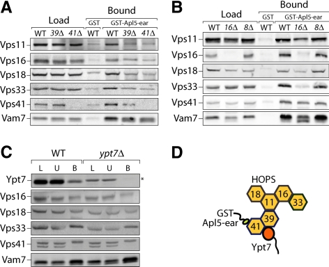
Adaptor protein complexes (APs) are evolutionarily conserved heterotetramers that couple cargo selection to the formation of highly curved membranes during vesicle budding. In Saccharomyces cerevisiae, AP-3 mediates vesicle traffic from the late Golgi to the vacuolar lysosome. The HOPS subunit Vps41 is one of the few proteins reported to have a specific role in AP-3 traffic, yet its function remains undefined. We now show that although the AP-3 delta subunit, Apl5, binds Vps41 directly, this interaction occurs preferentially within the context of the HOPS docking complex. Fluorescence microscopy indicates that Vps41 and other HOPS subunits do not detectably colocalize with AP-3 at the late Golgi or on post-Golgi (Sec7-negative) vesicles. Vps41 and HOPS do, however, transiently colocalize with AP-3 vesicles when these vesicles dock at the vacuole membrane. In cells with mutations in HOPS subunits or the vacuole SNARE Vam3, AP-3 shifts from the cytosol to a membrane fraction. Fluorescence microscopy suggests that this fraction consists of post-Golgi AP-3 vesicles that have failed to dock or fuse at the vacuole membrane. We propose that AP-3 remains associated with budded vesicles, interacts with Vps41 and HOPS upon vesicle docking at the vacuole, and finally dissociates during docking or fusion.


Figures

Figure 1.
Figure 1.
GST-Apl5-ear binds HOPS subunits. Pulldowns on GST-Apl5-ear resin were performed as described (see Materials and Methods) with ∼150 OD600 nm × ml of BY4742 yeast detergent lysate. Bounds proteins were eluted by incubating beads in 20 mM Tris-Cl, 20 mM glutathione, and 600 mM NaCl for 10 min at 4°C (top panel) or SDS loading buffer and heating at 95°C for 5 min (bottom panel). Twenty percent of the load (L) and 20% of the unbound fraction (U) relative to the bound fraction (B) were loaded, separated by SDS-PAGE, and analyzed by immunoblot. Asterisk (*) indicates nonspecific bands detected by the antibodies. The various forms of ALP are indicated: p, proALP; m, mature ALP; and s, soluble ALP. In wild-type cells, a large majority of ALP exists in the mature form.
Figure 2.
Figure 2.
GST-Apl5-ear interacts with the HOPS holocomplex. (A) Cell lysate from BJ3505 GFP-Vps39 was incubated for 1 h with GST-Apl5-ear glutathione resin. The resin was washed, and interacting proteins were eluted by a linear gradient of 0–0.7 M NaCl. Fractions were immunoblotted for the presence of HOPS. (B) Peak HOPS fractions were pooled, and 500 μl of the pooled peak was subjected to immunoprecipitation by α-GFP antibodies immobilized on Protein A-Sepharose. Note that the unbound material is depleted of all HOPS subunits but not of Vam7, which binds to AP-3 independently of HOPS. (C) The pooled HOPS peak was also subjected to size exclusion chromatography on a Superose 6 column. In calibration runs, blue dextran (2 MDa) eluted at fraction 17 (not shown), and thyroglobulin (670 kDa) eluted at fraction 26, as indicated. The exclusion limit of Superose 6 is Mr ∼4 × 10⁷.
Figure 3.
Figure 3.
An intact holocomplex is required for efficient Apl5 binding by HOPS subunits. Pulldowns on GST-Apl5-ear were performed using detergent lysate from BY4742 (WT) and the deletion mutants (A) vps39Δ and vps41Δ, (B) vps16Δ and vps8Δ, or (C) ypt7Δ as described for Figure 1. Interacting proteins were eluted by incubating beads twice in 20 mM Tris-Cl, pH 7.9, 20 mM glutathione, 600 mM NaCl, and 1% Triton X-100 for 10 min at 4°C. The load (L) and unbound (U) fractions correspond to 20% of the bound (B) fraction. Asterisk (*) indicates a nonspecific band detected by the Ypt7 antibody. Note that the Vps16 antibody also detects a nonspecific band that is enriched on the GST-Apl5 resin but not the GST resin, as seen in the vps16Δ eluates, resulting in a higher apparent Vps16 signal. (D) AP-3 preferentially binds Vps41 as part of the HOPS complex. The organization of the HOPS subunits is a composite model from yeast two-hybrid assays and immunoprecipitation from the Merz Lab (unpublished results) as well as published literature (Rieder et al., 1997; Wurmser et al., 2000). Although Vps41 interacts with the remaining HOPS subunits in the absence of Vps39 (unpublished results), the exact subunit interactions have not been mapped.
Figure 4.
Figure 4.
Purified GST-Apl5-ear binds to purified Vps41. Five micrograms of purified Vps41 or Vps33 was incubated with each of the GST-AP-ear constructs. After incubation at 4°C for 1 h, the beads were washed, and bound protein was eluted with SDS loading buffer. The load fraction corresponds to 2% of the bound fractions.
Figure 5.
Figure 5.
Apl5 transiently colocalizes with HOPS subunits at the vacuole membrane. Apl5 was C-terminally tagged with either GFP or mCherry. (A) Strains containing these Apl5 fusions were analyzed for ALP maturation using differential centrifugation (see Materials and Methods). (B) Strains containing Apl5-mCherry and a GFP-tagged HOPS subunit (Vps16-GFP, Vps33-GFP, GFP-Vps41, and GFP-Vps39) were grown to mid-log phase and analyzed by fluorescent microscopy. Arrows indicate sites of colocalization between Apl5 puncta and HOPS at the vacuole membrane. For the GFP-Vps39 time average (part B, bottom panels), both GFP and mCherry fluorescence were averaged across 10 frames (∼24 s). (C) Wild-type yeast expressing Apl5-GFP were labeled with FM4-64 to stain the vacuole membrane. For the 10-frame average, both GFP and FM4-64 channels were averaged across 10 frames (∼17 s). Bar, 2 μm. See Supplemental Movies 1–3.
Figure 6.
Figure 6.
Sec7-DsRed partially colocalizes with Apl5-GFP but not with GFP-HOPS subunits. Cells expressing Sec7-DsRed and (A) Apl5-GFP or (B) GFP-HOPS subunits were analyzed by fluorescence microscopy. Bar, 2 μm. See Supplemental Movies 4–7.
Figure 7.
Figure 7.
Deletion of docking and fusion factors results in AP-3 redistribution. Differential centrifugation of BY4742 (WT), ypt7Δ, vam3Δ, vps16Δ, vps41Δ, and vps8Δ mutant yeast lysates was performed as described in Materials and Methods. The load lane corresponds to 20% of the other fractions. A weak nonspecific band was detected by the Ypt7 antibody (*). For clarity, this band is indicated only for the ypt7Δ mutant.
Figure 8.
Figure 8.
Disruption of vacuole SNARE function or Vps41 deletion results in accumulation of post-Golgi AP-3 puncta. (A) Apl5-GFP localization was analyzed in VAM3 and vam3tsf cells at permissive (RT) and nonpermissive temperature (37°C). To determine the effect of mutations in (B) vps41Δ and (C) the dynamin homolog, vps1Δ, on AP-3 trafficking, these mutants were created in strains containing Apl5-GFP and Sec7-DsRed. For each figure, Apl5-GFP puncta were manually counted for each strain, with the results shown by box plot. Each box indicates the central 50% of the data (25th to 75th percentile), with the median denoted by a horizontal bar. Bars indicate the range. n = 50 cell profiles (A and B) or 100 cell profiles (C), using pooled data from two independent experiments. Significance values were calculated using the Mann-Whitney U test. Bar, 2 μm. See Supplemental Movies 8–10.
Figure 9.
Figure 9.
Working model for the AP-3 pathway in budding yeast. See text for discussion.

References

    1. Anand V. C., Daboussi L., Lorenz T. C., Payne G. S. Genome-wide analysis of AP-3 dependent protein transport in yeast. Mol. Biol. Cell. 2009;20:1592–1604. - PMC - PubMed
    1. Baggett J. J., Wendland B. Clathrin function in yeast endocytosis. Traffic. 2001;2:297–302. - PubMed
    1. Bonifacino J. S., Glick B. S. The mechanisms of vesicle budding and fusion. Cell. 2004;116:153–166. - PubMed
    1. Brett C. L., Plemel R. L., Vignali M., Fields S., Merz A. J. Efficient termination of vacuolar Rab GTPase signaling requires coordinated action by a GAP and a protein kinase. J. Cell Biol. 2008;182:1141–1151. - PMC - PubMed
    1. Cai H., Yu S., Menon S., Cai Y., Lazarova D., Fu C., Reinisch K., Hay J. C., Ferro-Novick S. TRAPPI tethers COPII vesicles by binding the coat subunit Sec23. Nature. 2007;445:941–944. - PubMed

Publication types

MeSH terms

Substances