Comparative Study
. 2009 Sep 9;29(36):11393-8.
doi: 10.1523/JNEUROSCI.2021-09.2009.

Abeta immunotherapy: intracerebral sequestration of Abeta by an anti-Abeta monoclonal antibody 266 with high affinity to soluble Abeta

Affiliations
Comparative Study

Abeta immunotherapy: intracerebral sequestration of Abeta by an anti-Abeta monoclonal antibody 266 with high affinity to soluble Abeta

Kaoru Yamada et al. J Neurosci. .

Abstract

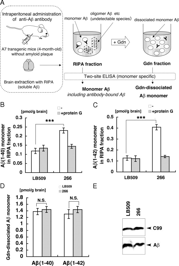
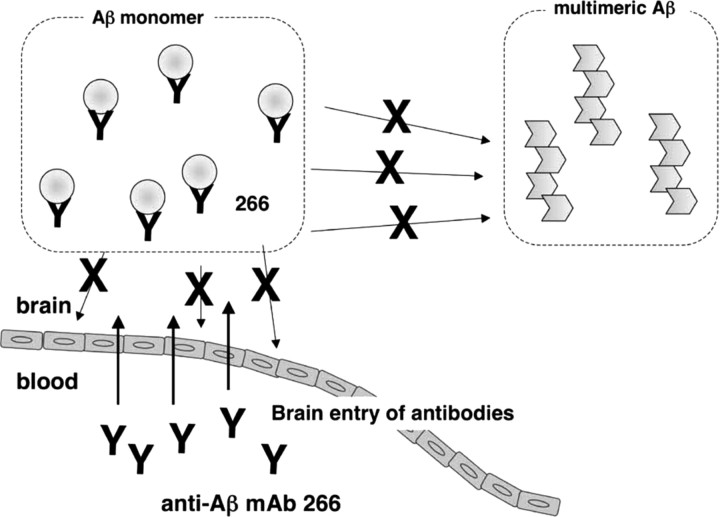
Amyloid beta (Abeta) immunotherapy is emerging as a promising disease-modifying therapy for Alzheimer's disease, although the precise mechanisms whereby anti-Abeta antibodies act against amyloid deposition and cognitive deficits remain elusive. To test the "peripheral sink" theory, which postulates that the effects of anti-Abeta antibodies in the systemic circulation are to promote the Abeta efflux from brain to blood, we studied the clearance of (125)I-Abeta(1-40) microinjected into mouse brains after intraperitoneal administration of an anti-Abeta monoclonal antibody 266. (125)I-Abeta(1-40) was rapidly eliminated from brains with a half-life of approximately 30 min in control mice, whereas 266 significantly retarded the elimination of Abeta, presumably due to formation of Abeta-antibody complex in brains. Administration of 266 to APP transgenic mice increased the levels of monomer Abeta species in an antibody-bound form, without affecting that of total Abeta. We propose a novel mechanism of Abeta immunotherapy by the class of anti-Abeta antibodies that preferentially bind soluble Abeta, i.e., intracerebral, rather than peripheral, sequestration of soluble, monomer form of Abeta, thereby preventing the accumulation of multimeric toxic Abeta species in brains.


Figures

Figure 1.
Figure 1.
Brain injection and clearance of 125I-Aβ1-40 after peripheral administration of anti-Aβ antibodies in mice. A, Time course of intraperitoneal administration of antibodies and 125I-Aβ injection. 125I-Aβ was injected into the cortex of mice brains 1, 24, or 120 h after antibody administration. The radioactivities of Aβ remaining in the brain were calculated as 100 − BEI (%) at 15, 30, 60, or 120 min after injection of 125I-Aβ. B, Time course of elimination of 125I-Aβ from brains 120 h after intraperitoneal administration of 10D5, 266, or LB509 as a control IgG. The means ± SEMs in three to eight independent assays are shown. **p < 0.01, ANOVA. C, The remaining 125I-Aβ at 60 min after microinjection of 125I-Aβ at 1, 24, or 120 h after intraperitoneal administration of 266 or LB509. The means ± SEMs in three to eight independent assays are shown. *p < 0.05, **p < 0.01, by Student's t test.
Figure 2.
Figure 2.
Formation of 125I-Aβ/antibody complex in mouse brains. A, Peripheral administration of antibodies 10D5, 266, or LB509 followed by intracerebral injection of 125I-Aβ and protein G precipitation of antibodies from brain extracts. 125I-Aβ was injected 5 d after intraperitoneal administration of antibodies. After extensive transcardiac perfusion with PBS, brains were removed and extracted with RIPA buffer and the resulting supernatants were precipitated by protein G agarose. The radioactivity derived from 125I-Aβ bound to the protein G-precipitated antibody as a percentage of total injected 125I-Aβ is shown. The means ± SEMs in three independent assays are shown. ***p < 0.001, ANOVA. B, Coinjection of 125I-Aβ with antibodies 10D5, 266, or LB509 (1 mg/ml) into brains. Apparent elimination rate constant (kapp,el %) of 125I-Aβ efflux is indicated as a percentage of that with LB509 (control antibody) as 100%. The means ± SEMs in three independent assays are shown. **p < 0.01, ANOVA.
Figure 3.
Figure 3.
Differential extraction of brains of APP transgenic mice following peripheral administration of anti-Aβ antibodies. A, Procedures of ELISA quantitation of Aβ in two sequentially prepared fractions from brains of APP transgenic mice. The level of “monomer Aβ” is defined as the concentration of Aβ in RIPA fraction detected by monomer-specific two-site ELISA, and “Gdn-dissociated soluble Aβ” as that of Aβ in the Gdn fraction. The levels of monomer Aβ1-40 (B) and Aβ1-42 (C) in the RIPA fractions of 4-month-old A7 mice 120 h after intraperitoneal administration of 266 or LB509 were quantitated by ELISA with (shaded) or without (blank) immunodepletion by protein G. The means ± SEMs in four independent assays are shown. ***p < 0.001, ANOVA. D, The levels of Gdn-dissociated monomer Aβ in the Gdn-fraction of the brains of 4-month-old A7 mice 120 h after intraperitoneal administration of 266 (shaded) or LB509 (blank) were quantitated by ELISA. The means ± SEMs in four independent assays are shown. E, The total Aβ and its precursor C99 in the RIPA fractions were analyzed by immunoblotting with 82E1 after immunoprecipitation with the same antibody.
Figure 4.
Figure 4.
Possible mechanism of action of anti-Aβ mAb 266 in passive Aβ immunotherapy. 266 enters the brain parenchyma and sequesters Aβ chiefly in a monomeric state, thereby inhibiting further multimerization of Aβ and neurotoxicity.

References

    1. Baba M, Nakajo S, Tu PH, Tomita T, Nakaya K, Lee VM, Trojanowski JQ, Iwatsubo T. Aggregation of α-synuclein in Lewy bodies of sporadic Parkinson's disease and dementia with Lewy bodies. Am J Pathol. 1998;152:879–884. - PMC - PubMed
    1. Bacskai BJ, Kajdasz ST, McLellan ME, Games D, Seubert P, Schenk D, Hyman BT. Non-Fc-mediated mechanisms are involved in clearance of amyloid-β in vivo by immunotherapy. J Neurosci. 2002;22:7873–7878. - PMC - PubMed
    1. Bard F, Cannon C, Barbour R, Burke RL, Games D, Grajeda H, Guido T, Hu K, Huang J, Johnson-Wood K, Khan K, Kholodenko D, Lee M, Lieberburg I, Motter R, Nguyen M, Soriano F, Vasquez N, Weiss K, Welch B. Peripherally administered antibodies against amyloid β-peptide enter the central nervous system and reduce pathology in a mouse model of Alzheimer disease. Nat Med. 2000;6:916–919. - PubMed
    1. DeMattos RB, Bales KR, Cummins DJ, Dodart JC, Paul SM, Holtzman DM. Peripheral anti-Aβ antibody alters CNS and plasma Aβ clearance and decreases brain Aβ burden in a mouse model of Alzheimer's disease. Proc Natl Acad Sci U S A. 2001;98:8850–8855. - PMC - PubMed
    1. DeMattos RB, Bales KR, Cummins DJ, Paul SM, Holtzman DM. Brain to plasma amyloid-β efflux: a measure of brain amyloid burden in a mouse model of Alzheimer's disease. Science. 2002a;295:2264–2267. - PubMed

Publication types

MeSH terms

Substances