. 2009 Dec;86(6):1275-83.
doi: 10.1189/jlb.0209107. Epub 2009 Sep 9.

The sympathetic nervous system modulates CD4(+)FoxP3(+) regulatory T cells via a TGF-beta-dependent mechanism

Affiliations

The sympathetic nervous system modulates CD4(+)FoxP3(+) regulatory T cells via a TGF-beta-dependent mechanism

Sourojit Bhowmick et al. J Leukoc Biol. 2009 Dec.

Abstract

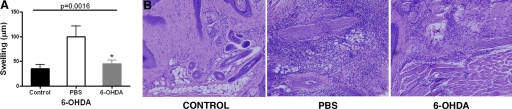
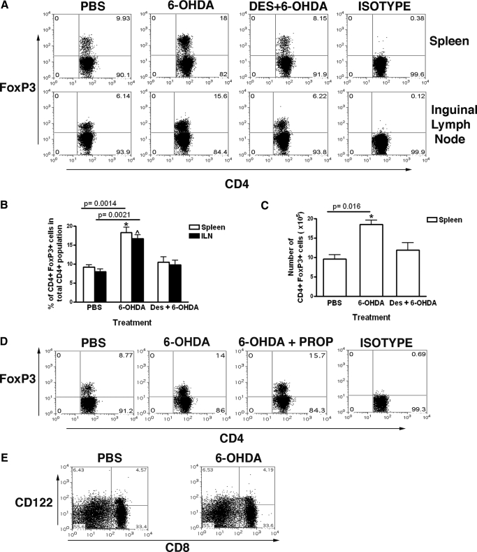
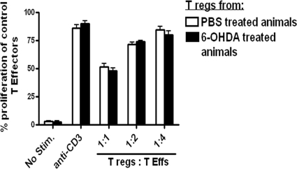
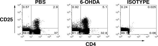
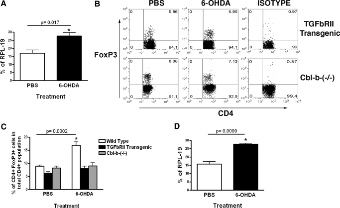
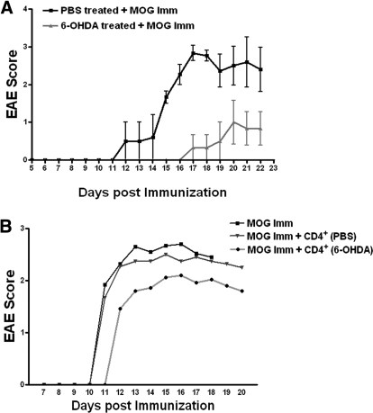
CD4(+)FoxP3(+) Tregs are essential mediators of the peripheral immune response to self-antigens. Accordingly, the homeostatic regulation of Treg activity and number would impact on the immune response to both self- and non-self antigens. Because the sympathetic nervous system (SNS) interacts chemically and physically with the central and peripheral immune system and exerts a direct influence on antigen-presenting cells and effector lymphocytes, we have investigated the effect of chemical ablation of the SNS on the number and function of peripheral Treg. Removal of murine peripheral sympathetic innervation by 6-hydroxydopamine induced an increase in splenic and lymph node CD4(+)FoxP3(+) Tregs by a TGF-beta-dependent mechanism. Further, this increase in Tregs coincides with an inhibition of the induction of experimental autoimmune encephalomyelitis. Our results demonstrate that the SNS is an important contributor to the maintenance of peripheral Treg and TGF-beta acts as a bridge between the immune system and the nervous system. Neurological events mediated by the SNS, such as a stress response, may affect the number of T cells that regulate an immune response. Additionally, targeting Tregs via the SNS may be a novel approach to the prevention or treatment of autoimmune diseases.


Figures

Figure 1.
Figure 1.
Abrogation of DTH reaction in 6-OHDA-treated, immunized mice. Mice were immunized with MOG/CFA 2 days after receiving i.p. dose of 6-OHDA (200 mg/kg). Control groups did not receive any immunization. Seven days after immunizing, one footpad was challenged with the MOG and the other footpad with vehicle only. (A) The thickness of footpads measured 24 h after challenge. Bar graph represents the mean ± SE footpad swelling of 6 mice/group, 2 experiments. (B) Histology of the footpad 24 h after challenge with MOG from immunized mice without challenge (Control), immunized mice that received an i.p. injection of PBS or 6-OHDA 2 days before immunization and then challenged. Mice showed perivascular infiltration of cells in the PBS or 6-OHDA-treated groups. Magnification ×20.
Figure 2.
Figure 2.
Peripheral chemical sympathectomy by 6-OHDA induces an increase in the number of Tregs. (A) Representative dot plots show flow cytometric analysis of mouse (C57BL/6) spleen and inguinal lymph node cells at 48-h post injection of PBS, 6-OHDA, or desipramine and 6-OHDA. Plots represent gated CD4 T cells. The values in the upper-right quadrant represent CD4+ FoxP3+ T cells as a percentage of total CD4 T cells. (B) Bar graph shows the mean results (n=12–15). (C) Bar graph showing the absolute number of CD4+ FoxP3+ Tregs from the above experiment. Numbers were calculated from flow cytometry data from the spleen. (D) Propranolol fails to prevent the increase in Tregs by chemical sympathectomy: representative dot plots show flow cytometric analysis of mouse (C57BL/6) spleen and inguinal lymph node cells at 48 h postinjection of PBS, 6-OHDA, or 6-OHDA followed by propranolol. Plots represent gated CD4 T cells. The values in the upper-right quadrant represent CD4+ FoxP3+ T cells as a percentage of total CD4 T cells. (E) 6-OHDA does not effect the number of total CD8+ cells or CD8+ CD122+ cells. Representative dot plots show flow cytometric analysis of mouse (C57BL/6) spleen cells at 48 h postinjection of PBS or 6-OHDA. Cells are gated for CD3+. The values in the upper right quadrant represent CD8+ CD122+ T cells.
Figure 3.
Figure 3.
Tregs from 6-OHDA-treated mice are functionally similar to Tregs from PBS-treated mice. In vitro Treg functional assay: CD4+CD25 T cells (T effs) from the spleen of naïve C57BL/6 were labeled with CFSE, in each group. CFSE-labeled responder T effs did not proliferate in unstimulated conditions (No Stim.) but did proliferate in the presence of soluble anti-CD3 as demonstrated by the dilution of CFSE in the stimulated group. Tregs cocultured with T effs and antigen-presenting cells (live splenic DCs) in the presence of anti-CD3, as described, suppress the proliferation of responder T eff cells as quantified by CFSE dilution. Tregs were isolated from control or 6-OHDA-treated animals and cocultured with naïve T effs at a 1:1, 1:2, 1:4 Treg-to-T eff ratio.
Figure 4.
Figure 4.
6-OHDA induced sympathectomy does not change the frequency of total CD4+ T cells. C57BL/6 mice were treated with 6-OHDA, while control mice received PBS. Forty-eight hours after injection, total spleen cells were stained. Representative FACS dot plots demonstrate the percentage of CD4+ CD25+ T cells in the spleen, 48 h after treatment. Plots represent a total lymphocyte gate. The values represent different cell types, as a percentage of total lymphocytes.
Figure 5.
Figure 5.
6-OHDA-induced increase in Tregs is mediated by TGF-β. (A) TGF-β mRNA expression in PBS or 6-OHDA-treated C57BL/6 mice, assessed by qRT-PCR from total spleen cells taken 48 h after injection. Bar graph shows TGF-β mRNA expression as a percentage of RPL-19 (a housekeeping gene) expression. (B) Representative FACS dot plots demonstrating the percentage of CD4+ FoxP3+ Tregs in the spleens of PBS-treated or 6-OHDA treated TGFbRII transgenic and Cbl-b-(−/−) mouse, 48 h after treatment. Plots represent gated CD4 T cells. The values in the upper right quadrant represent CD4+ FoxP3+ T cells as a percentage of total CD4 T cells. (C) Bar graph shows the mean result (n=10). (D) TGF-β mRNA expression before and after 6-OHDA induced peripheral sympathectomy in TGFbRII mice qRT-PCR from total spleen cells taken 48 h after injection. Bar graph shows TGF-β mRNA expression as percentage of RPL-19 expression.
Figure 6.
Figure 6.
6-OHDA-induced peripheral chemical sympathectomy prevents EAE. (A) Female C57BL/6 mice, 7 to 8 wk old were injected with 6-OHDA, and then, 48 h later were immunized with MOG, as described previously. Clinical scores of vehicle-treated mice (light shade) and mice treated with 6-OHDA (dark shade) (0, normal; 1, limp tail; 2, paraparesis with a clumsy gait; 3, hind limb paralysis; 4, quadriplegia; 5, death) were assessed each day. Data represent mean scores (n=10, vehicle and; n=12, 6-OHDA) ± SE. (B) Mice were immunized with MOG peptide and received 1 × 10⁶ CD4+ T cells (i.v.) from PBS or 6-OHDA-treated mice immediately after immunization. Clinical scores of uninjected (black), PBS-CD4+ T cells-injected (—▾—) and 6-OHDA- CD4+ T cells-injected (light shade) mice, assessed each day. Data represent mean scores (n=10, uninjected; n=10, PBS-CD4+ T cells injected; and n=12, 6-OHDA-CD4+ T cells injected).

Comment in

References

    1. Hogquist K A, Baldwin T A, Jameson S C. Central tolerance: learning self-control in the thymus. Nat Rev Immunol. 2005;5:772–782. - PubMed
    1. Goodnow C C, Sprent J, Fazekas de St Groth B, Vinuesa C G. Cellular and genetic mechanisms of self tolerance and autoimmunity. Nature. 2005;435:590–597. - PubMed
    1. Fontenot J D, Gavin M A, Rudensky A Y. Foxp3 programs the development and function of CD4+CD25+ regulatory T cells. Nat Immunol. 2003;4:330–336. - PubMed
    1. Sakaguchi S. Naturally arising Foxp3-expressing CD25+CD4+ regulatory T cells in immunological tolerance to self and non-self. Nat Immunol. 2005;6:345–352. - PubMed
    1. Annacker O, Pimenta-Araujo R, Burlen-Defranoux O, Barbosa T C, Cumano A, Bandeira A. CD25+ CD4+ T cells regulate the expansion of peripheral CD4 T cells through the production of IL-10. J Immunol. 2001;166:3008–3018. - PubMed

Publication types

MeSH terms