. 2009 Nov;297(5):C1071-81.
doi: 10.1152/ajpcell.00284.2009. Epub 2009 Sep 9.

Loss of the apical V-ATPase a-subunit VHA-6 prevents acidification of the intestinal lumen during a rhythmic behavior in C. elegans

Affiliations

Loss of the apical V-ATPase a-subunit VHA-6 prevents acidification of the intestinal lumen during a rhythmic behavior in C. elegans

Erik Allman et al. Am J Physiol Cell Physiol. 2009 Nov.

Abstract

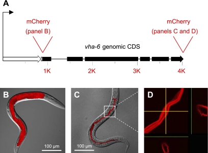
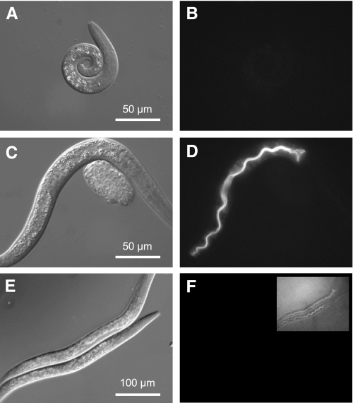
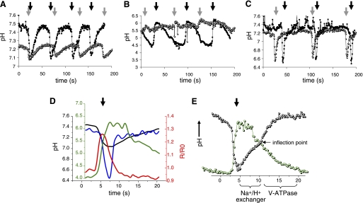
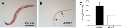
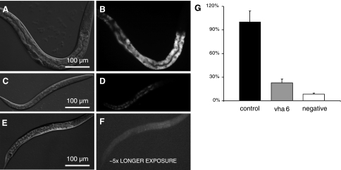
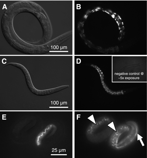
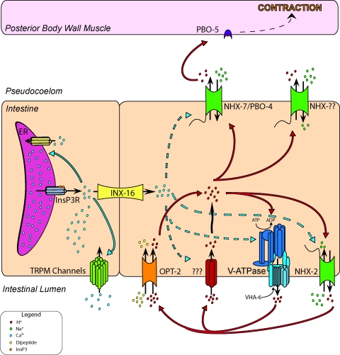
In Caenorhabditis elegans, oscillations of intestinal pH contribute to the rhythmic defecation behavior, but the acid-base transport mechanisms that facilitate proton movement are not well understood. Here, we demonstrate that VHA-6, an intestine-specific a-subunit of the H(+)-K(+)-ATPase complex (V-ATPase), resides in the apical membrane of the intestinal epithelial cells and is required for luminal acidification. Disruption of the vha-6 gene led to early developmental arrest; the arrest phenotype could be complemented by expression of a fluorescently labeled vha-6 transgene. To study the contribution of vha-6 to pH homeostasis in larval worms, we used a partial reduction of function through postembryonic single-generation RNA interference. We demonstrate that the inability to fully acidify the intestinal lumen coincides with a defect in pH recovery of the intestinal epithelial cells, suggesting that VHA-6 is essential for proton pumping following defecation. Moreover, intestinal dipeptide accumulation and fat storage are compromised by the loss of VHA-6, suggesting that luminal acidification promotes nutrient uptake in worms, as well as in mammals. Since acidified intracellular vesicles and autofluorescent storage granules are indistinguishable between the vha-6 mutant and controls, it is likely that the nutrient-restricted phenotype is due to a loss of plasma membrane V-ATPase activity specifically. These data establish a simple genetic model for proton pump-driven acidification. Since defecation occurs at 45-s intervals in worms, this model represents an opportunity to study acute regulation of V-ATPase activity on a short time scale and may be useful in the study of alternative treatments for acid-peptic disorders.


Figures

Fig. 1.
Fig. 1.
Expression and localization of the vha-6 gene product in transgenic worms. A: vha-6 promoter (open arrow) and genomic coding region (filled arrow) and sites where mCherry cDNA was inserted to create transcriptional and translational fusions are depicted to scale. B: transcriptional fusion to the 1,023-nucleotide promoter results in mCherry expression restricted exclusively to intestinal epithelia of the worm at the L4 stage. C: translational fusion that included the promoter and the genomic coding region fused in-frame to the mCherry cDNA results in protein targeting to the apical membrane of intestinal epithelial cells of the L4 hermaphrodite. B and C are overlays of confocal microscopy maximum projection images onto their corresponding differential interference contrast (DIC) images. D: magnification of the portion of C enclosed in the white box and display of cross-sectional images in y- and z-planes (yellow arrows) clearly demonstrate that the fusion protein lies at or near the apical surface. CDS, coding region.
Fig. 2.
Fig. 2.
Analysis of vha-6 mutant and RNA interference (RNAi) phenotypes. A, C, and E are DIC contrast images; B, D, and F are fluorescent images. Homozygous vha-6(ok1825) deletion mutants (courtesy of the C. elegans Gene Knockout Consortium) arrested development as L1 larva, but an extrachromosomal array coding for a Pvha-6::VHA-6::mCherry translational fusion could complement the mutation and restore normal growth. To illustrate this, we examined brethren from the rescued line where mosaic loss of the rescue construct at 15°C resulted in worms that lacked fluorescence and arrested development as L1 larvae (A and B; 48-h posthatch L1 arrest), whereas the presence of the construct in age-matched transgenic siblings restored normal development (C and D; 48-h posthatch L2/L3). Single-generation treatment of wild-type worms with vha-6 RNAi for 3 days, starting as a recently hatched L1 larva, reduced VHA-6::mCherry expression to nearly undetectable levels (F; inset is 10-fold longer exposure). RNAi-treated worms exhibited a progressive slowing of growth, such that they had generally reached the L3/L4 stage by 72 h and often never reached reproductive maturity (E). These phenotypes are consistent with a requirement for VHA-6 activity for growth and maturation.
Fig. 3.
Fig. 3.
Analysis of intestinal pH dynamics in free-range worms. Representative traces from single 72-h posthatch animals show intracellular pH oscillations (A), luminal pH oscillations (B), and basolateral, extracellular pH oscillations (C) in control (▪) and vha-6(RNAi) (○) worms imaged over 200 s while moving freely on bacteria-seeded plates. pBoc is signified by dark arrows (control) and gray arrows (vha-6) to show time intervals for a complete defecation period in the worms. Intracellular and luminal data (A and B) were collected from worms in which RNAi feeding was initiated upon hatching. Because the extracellular pH biosensor is not expressed well in larva, to allow development to adulthood, we collected basolateral data (C) from worms exposed to RNAi at the L3 stage. Their older age likely accounts for their somewhat extended defecation period. Average traces of single cycles from multiple worms are shown in the supplemental data, and statistics are provided in Table 1. D: representation of intestinal pH [intracellular pH (pHi, black trace), luminal pH (green trace), and basolateral, extracellular pH (blue trace)] and calcium oscillations (red trace) during defecation. All traces are plotted to the axis of the same color, except the basolateral trace, which is plotted to the black axis, the same as pHi. Individual traces have been normalized to the execution of pBoc at the start of defecation but were collected from separate worms and are not intended to depict exact temporal or causal relationships. R/R0, ratio normalized to time 0. E: biphasic kinetics of pHi recovery suggest that 2 ion transport processes function in sequence (black trace). The first phase is predicted to occur through Na+/H+ exchange, as indicated, which facilitates recovery from acidification in most cell types. This phase exhibits pH dependence and an apparent plateau near the inflection point. Whether these protons are exported from the cell across the basolateral or the apical membrane is unknown, although luminal pH is relatively stable during this first phase of cytoplasmic recovery (green trace). The second phase is clearly noted after the inflection point in the pHi trace. During this phase, luminal pH begins to decrease as a result of proton pumping at the apical membrane, and the cell is driven to become more alkaline. Traces were not extracted from a single animal, and the relationship between cytoplasmic and luminal pH is intended to be representative, rather than absolute. Scale has been optimized for comparison (for actual values see Table 1)
Fig. 4.
Fig. 4.
Analysis of fat storage. Images of Oil Red O-stained lipid droplets suggest a severe reduction of fat mass in small, adult vha-6(RNAi) worms (B) compared with adult control worms (A). Quantification of staining intensity (C) confirmed a large reduction of whole body fat mass after vha-6 targeting (n = 20, P < 0.01); background staining was negligible. Arrows in B denote mature oocytes, which retain Oil Red O staining, even in the absence of VHA-6, and contribute to the total body fat mass shown in C.
Fig. 5.
Fig. 5.
Analysis of nutrient uptake. DIC (A, C, and E) and corresponding fluorescent (B, D, and F) images of control (A and B) and vha-6(RNAi) (C and D) worms that were, after 48 h of RNAi, cultured overnight in liquid medium containing a bacterial food source and AMCA-labeled Ala-Lys dipeptide. Negative controls (wild-type larva, no dipeptide; E and F) displayed slight fluorescence, despite longer exposure. Quantification of staining intensity (G) confirms a significant reduction of dipeptide uptake (n = 10, P < 0.01).
Fig. 6.
Fig. 6.
Analysis of acidic gut granules in adult worms. LysoTracker Red was used to stain acidic gut granules in 3-day-old control (A and B) and vha-6(RNAi) worms (C and D). Staining intensity varied along the length of the intestine, with posterior intestinal cells displaying the brightest signal. Control and vha-6(RNAi) worms stained similarly, despite the obvious difference in developmental age (L2 vs. young adult); developmentally matched worms similarly showed no obvious differences in the formation of acidic granules. Inset in D shows a negative control (without LysoTracker Red) that was exposed 5 times as long, demonstrating lack of background. For examination of autofluorescent gut granules, 3-fold embryos from the mosaic vha-6(ok1825) Pvha-6::VHA-6::mCherry rescue line were imaged at 555-nm excitation and 590-nm emission for transgene expression (E) and at 340-nm excitation and 535-nm emission for autofluorescence (F). Embryo at right is expressing mCherry rescue marker at the apical membrane of the lumen, which fluoresces red (E), as well as a muscle Pmyo-3::GFP marker used to track the transgene (arrow in F). Embryo at left lacks both transgenes, yet it accumulates UV-excitable autofluorescent gut granules (arrowheads in F) to the same extent as its transgenic sibling, indicating that successful biogenesis of these acidic intracellular storage compartments occurs in the complete absence of vha-6 expression.
Fig. 7.
Fig. 7.
Schematic of rhythmic intestinal electrolyte transport during defecation in worms. Resting pH of the intestinal cell and the pseudocoelomic fluid that bathes the cell's basolateral surface is near a physiological norm of 7.4. Lumen of the intestine is acidic, with pH closer to 4. Cell-autonomous oscillatory calcium signaling involving activation of inositol 1,4,5-trisphosphate receptor (InsP3R) underlies the defecation pacemaker and triggers calcium inflow through the transient receptor potential melastatin channels GTL-1 and GON-2. A calcium wave propagates through the intestine, moving between adjacent intestinal cells through the gap junction protein INX-16. Elevations in calcium appear to cause the apical membrane to become permeable to protons, which enter the cell through an unknown mechanism. As protons flow down the transmembrane gradient, the cell acidifies and the lumen alkalinizes. In response to acidification and, perhaps, calcium as well, the Na+/H+ exchanger PBO-4/NHX-7 extrudes protons across the basolateral membrane. Protons trigger the first muscle contraction (pBoc) in the defecation motor program by binding to the muscle cell receptor PBO-5. Calcium returns to baseline levels in part through the activity of SCA-1 (SERCA), the endoplasmic reticular (ER) membrane ATPase, and pHi gradually recovers from acidification. This process likely involves Na+-H+ antiport and V-ATPase activities (see Fig. 3) but does not require NHX-7, which acts in a signaling, rather than a homeostatic, pathway. Eventually, luminal pH reaches ∼4, at which point the cytoplasm begins to acidify slightly. We hypothesize that this occurs as a result of metabolic processes that result in proton accumulation in the cell and inability of VHA-6 to export these protons, because the pH gradient has become too steep or because of some unknown regulatory pathway involving perhaps trafficking or subunit disassembly. In most acid-secreting mammalian systems, proton pumping requires the activity of apical K+ and Cl channels (such as KCNE2 and CFTR); whether this holds true in worms as well is under investigation. Finally, protons that have been pumped into the lumen work to allow nutrient absorption though proton-coupled uptake pathways such as the H+-dipeptide symporter OPT-2. This is a representative, rather than an exhaustive, depiction of the transporters that function in defecation.

References

    1. Al-Awqati Q. Plasticity in epithelial polarity of renal intercalated cells: targeting of the H+-ATPase and band 3. Am J Physiol Cell Physiol 270: C1571–C1580, 1996 - PubMed
    1. Ashrafi K, Chang FY, Watts JL, Fraser AG, Kamath RS, Ahringer J, Ruvkun G. Genome-wide RNAi analysis of Caenorhabditis elegans fat regulatory genes. Nature 421: 268–272, 2003 - PubMed
    1. Beg AA, Ernstrom GG, Nix P, Davis MW, Jorgensen EM. Protons act as a transmitter for muscle contraction in C. elegans. Cell: 149–160, 2008 - PMC - PubMed
    1. Beyenbach KW, Wieczorek H. The V-type H+ ATPase: molecular structure and function, physiological roles and regulation. J Exp Biol 209: 577–589, 2006 - PubMed
    1. Blair HC, Teitelbaum SL, Ghiselli R, Gluck S. Osteoclastic bone resorption by a polarized vacuolar proton pump. Science 245: 855–857, 1989 - PubMed

Publication types

MeSH terms

Substances

LinkOut - more resources