. 2010 Jan;58(1):17-28.
doi: 10.1369/jhc.2009.953604. Epub 2009 Sep 9.

Indoleamine 2,3-dioxygenase tissue distribution and cellular localization in mice: implications for its biological functions

Affiliations

Indoleamine 2,3-dioxygenase tissue distribution and cellular localization in mice: implications for its biological functions

Xiangchen Dai et al. J Histochem Cytochem. 2010 Jan.

Abstract

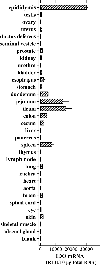
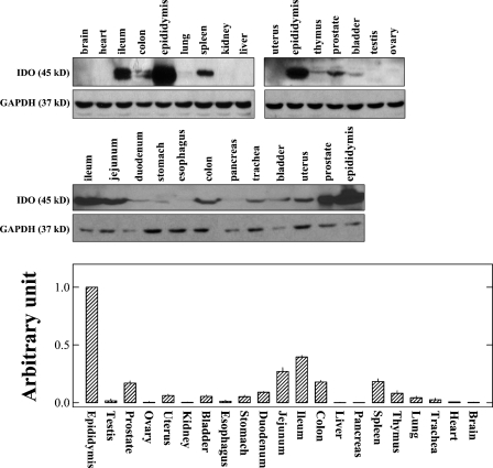
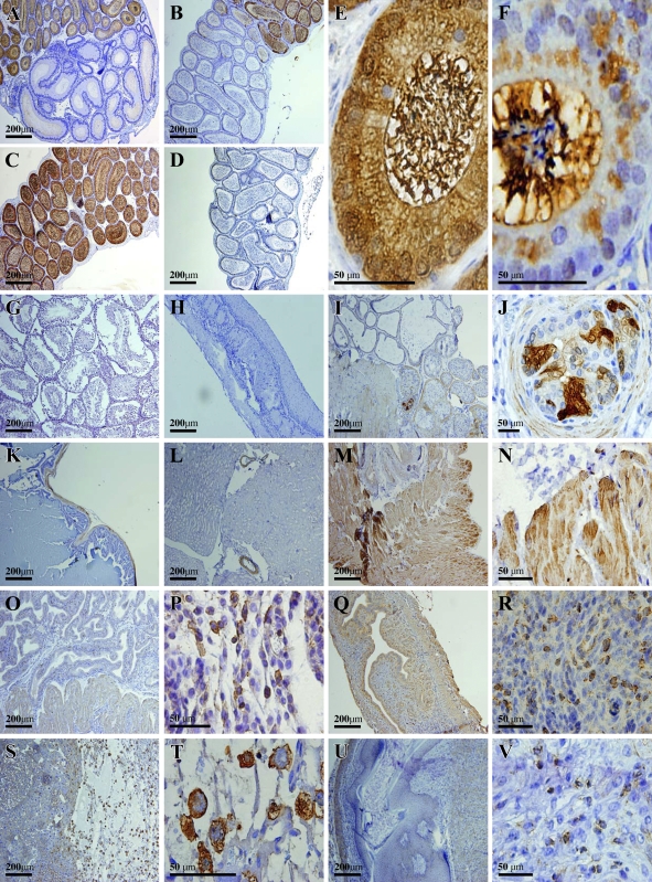
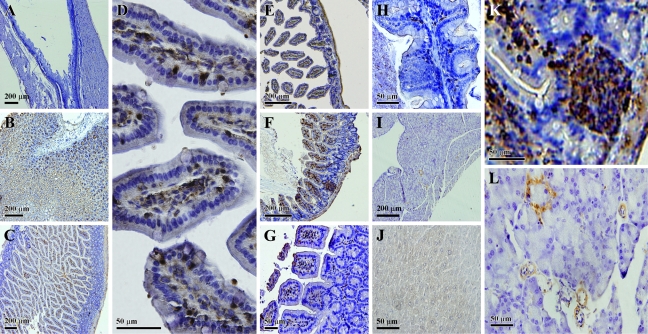
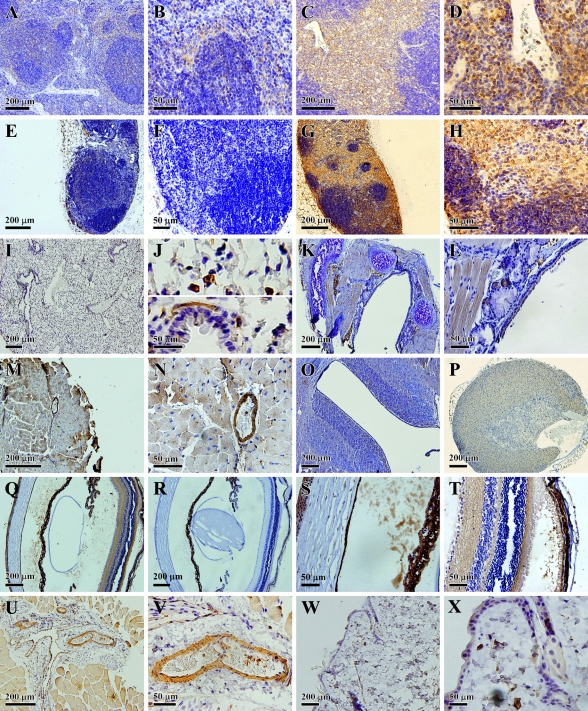
Earlier studies have suggested that indoleamine 2,3-dioxygenase (IDO) has a wide tissue distribution in mammals. However, detailed information on its cellular localization and also the levels of expression in various tissues is still scarce. In the present study, we sought to determine the cellular localization of IDO and also to quantify the level of its expression in various mouse tissues by using the branched DNA signal amplification assay, Western blotting, and immunohistochemical staining. The highest levels of constitutive IDO expression were found to be selectively present in the caput of epididymis, except for its initial segment. IDO expression was also detected inside the luminal compartment and even in the stereocilia within this region. In the prostate, high levels of IDO were selectively expressed in the capsular cells. In addition, high levels of IDO expression were also selectively detected in certain types of cells in the placenta, spleen, thymus, lung, and digestive tract. Notably, the morphological features of most of the positively stained cells in these organs closely resembled those of antigen-presenting cells. Based on the tissue distribution and cellular localization characteristics of IDO, it is hypothesized that its expression may serve two main functions: one is to deplete tryptophan in an enclosed microenvironment (such as in the epididymal duct lumen) to prevent bacterial or viral infection, and the other is to produce bioactive tryptophan catabolites that would serve to suppress T-cell-mediated immune responses against self-antigens, fetal antigens, or allogeneic antigens, in different situations.


Figures

Figure 1
Figure 1
Levels of indoleamine 2,3-dioxygenase (IDO) mRNA in various mouse tissues. The procedures for the collection of tissues and for the branched DNA signal amplification assay are described in detail in Materials and Methods. Each value is the mean ± SD of triplicate determinations. Two separate experiments were conducted, and similar data were obtained (one representative data set is shown).
Figure 2
Figure 2
Levels of IDO protein in various mouse tissues determined by Western blot analysis. The upper panel shows the representative IDO bands for different tissue samples detected by Western blotting, and the lower panel shows the quantitative comparison of IDO protein levels in different tissues based on densitometry measurements. The procedures for the collection of tissues and for the Western blotting analysis are described in detail in Materials and Methods. Note that for most tissues, the analysis was conducted multiple times. In each experiment, the proteins from pooled epididymis tissue were included as a control for comparison. The relative levels of IDO protein in each tissue are expressed as a relative unit that considers the IDO protein level in epidydymis to be 100%. Each value is the mean ± SD of at least three experiments.
Figure 3
Figure 3
Immunohistochemical localization of IDO protein in the mouse genitourinary system. (A) Initial and second segments of the caput of epididymis. (B) Last segment of the caput and corpus of epididymis. (C) Caput of epididymis (not including the initial segment). (D) Cauda of epididymis. (E) High magnification showing the stained cells in the caput of epididymis. (F) High magnification of stained cells in the caput of epididymis. Note that in this experiment, incubation of the tissue slides with DAB lasted for a shorter length of time than usual. (G) Testis; (H) ductus deferens; (I,J) prostate; (K) seminal vesicle; (L) kidney; (M,N) bladder; (O,P) uterus in the secretory phase; (Q,R) uterus (9th day of pregnancy); (S,T) placenta; (U,V) ovary.
Figure 4
Figure 4
Immunohistochemical localization of the IDO protein in the mouse digestive system. (A) Esophagus; (B) stomach; (C,D) low and high magnification, respectively, of stained cells in the duodenum; (E) jejunum; (F) ileum; (G) colon; (H) cecum; (I) pancreas; (J) liver; (K) high magnification of stained cells in the ileum; (L) high magnification of stained cells in the pancreas.
Figure 5
Figure 5
Immunohistochemical localization of IDO protein in other mouse systems. (A,B) Low and high magnification, respectively, of stained cells in the spleen. (C,D) Low and high magnification, respectively, of stained cells in the thymus. (E,F) Low and high magnification, respectively, of an axillary lymph node. (G,H) Low and high magnification, respectively, of stained cells in a jugular lymph node. (I,J) Low and high magnification, respectively, of stained cells in the lung. (K) Trachea. (L) Thyroid. (M,N) Low and high magnification, respectively, of stained cells in the heart. (O) Brain. (P) Adrenal gland. (Q) Low magnification of stained cells in the eye. (R) Low magnification of cells in the eye with negative staining. (S,T) High magnification of stained cells in the eye. (U,V) Low and high magnification, respectively, of stained cells in the skeletal muscle. (W,X) Low and high magnification, respectively, of stained cells in the skin.

References

    1. Bauer TM, Jiga LP, Chuang JJ, Randazzo M, Opelz G, Terness P (2005) Studying the immunosuppressive role of indoleamine 2,3-dioxygenase: tryptophan metabolites suppress rat allogeneic T-cell responses in vitro and in vivo. Transpl Int 18:95–100 - PubMed
    1. Beutelspacher SC, Pillai R, Watson MP, Tan PH, Tsang J, McClure MO, George AJ, et al. (2006) Function of indoleamine 2,3-dioxygenase in corneal allograft rejection and prolongation of allograft survival by over-expression. Eur J Immunol 36:690–700 - PubMed
    1. Britan A, Maffre V, Tone S, Drevet JR (2006) Quantitative and spatial differences in the expression of tryptophan-metabolizing enzymes in mouse epididymis. Cell Tissue Res 324:301–310 - PubMed
    1. Chiarugi A, Rapizzi E, Moroni F, Moroni F (1999) The kynurenine metabolic pathway in the eye: studies on 3-hydroxykynurenine, a putative cataractogenic compound. FEBS Lett 453:197–200 - PubMed
    1. Dai X, Zhu BT (2009) Suppression of T-cell response and prolongation of allograft survival in a rat model by tryptophan catabolites. Eur J Pharmacol 606:225–232 - PubMed

Publication types

MeSH terms