. 2009 Sep 9;4(9):e6919.
doi: 10.1371/journal.pone.0006919.

The G-quadruplex ligand telomestatin impairs binding of topoisomerase IIIalpha to G-quadruplex-forming oligonucleotides and uncaps telomeres in ALT cells

Affiliations

The G-quadruplex ligand telomestatin impairs binding of topoisomerase IIIalpha to G-quadruplex-forming oligonucleotides and uncaps telomeres in ALT cells

Nassima Temime-Smaali et al. PLoS One. .

Abstract

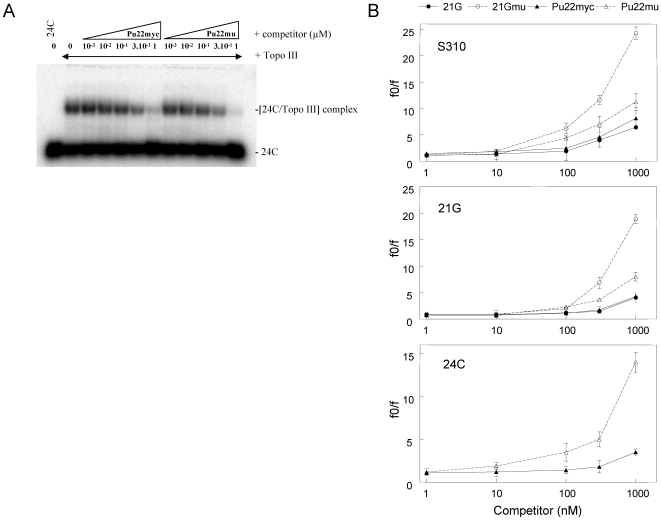
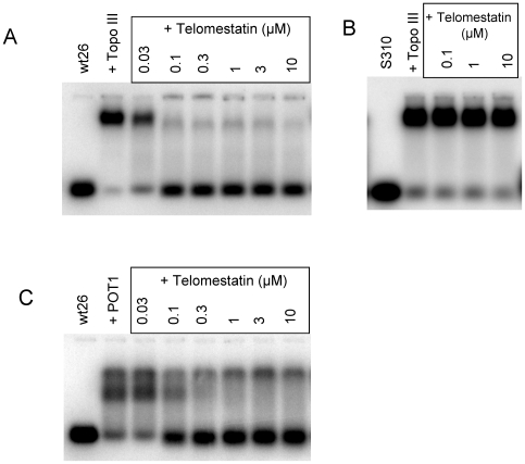
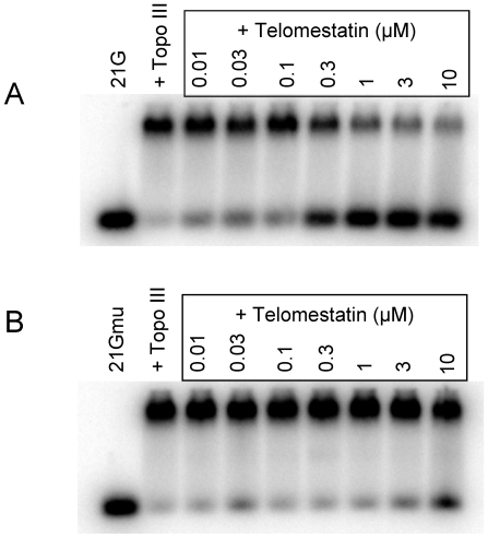
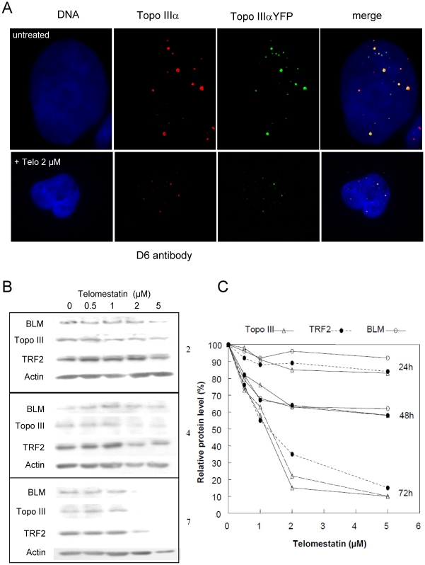
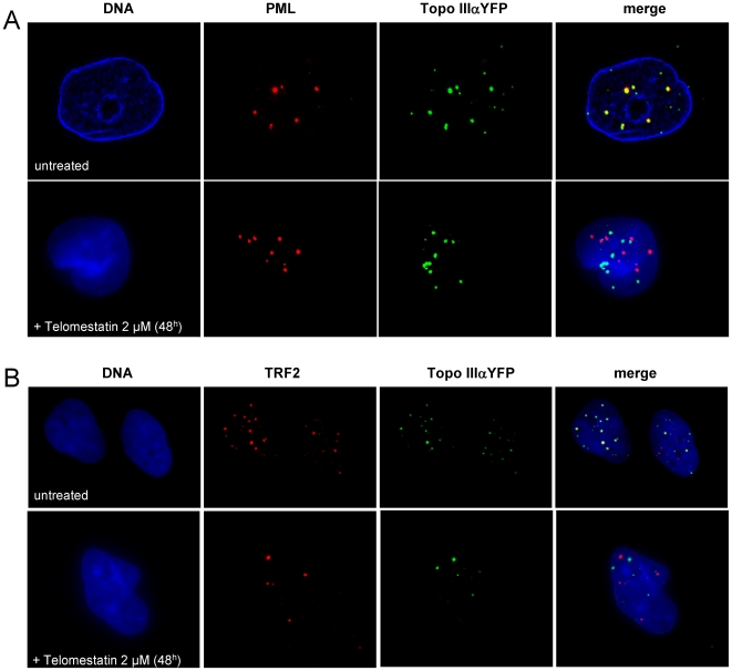
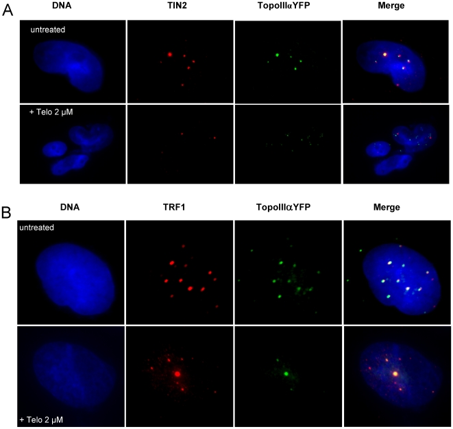
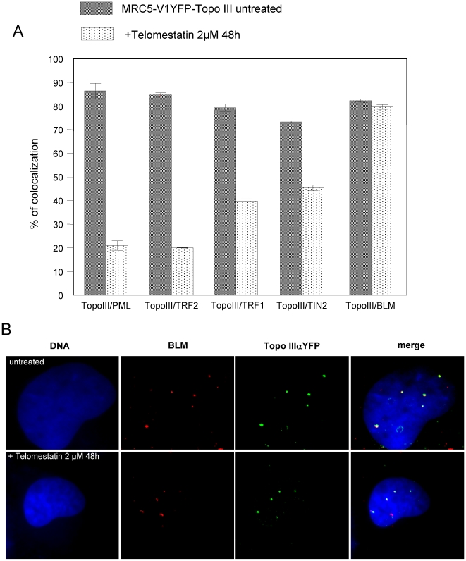
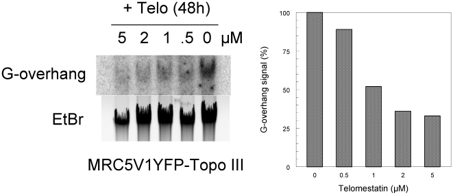
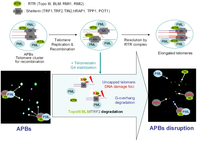
In Alternative Lengthening of Telomeres (ALT) cell lines, specific nuclear bodies called APBs (ALT-associated PML bodies) concentrate telomeric DNA, shelterin components and recombination factors associated with telomere recombination. Topoisomerase IIIalpha (Topo III) is an essential telomeric-associated factor in ALT cells. We show here that the binding of Topo III to telomeric G-overhang is modulated by G-quadruplex formation. Topo III binding to G-quadruplex-forming oligonucleotides was strongly inhibited by telomestatin, a potent and specific G-quadruplex ligand. In ALT cells, telomestatin treatment resulted in the depletion of the Topo III/BLM/TRF2 complex and the disruption of APBs and led to the segregation of PML, shelterin components and Topo III. Interestingly, a DNA damage response was observed at telomeres in telomestatin-treated cells. These data indicate the importance of G-quadruplex stabilization during telomere maintenance in ALT cells. The function of TRF2/Topo III/BLM in the resolution of replication intermediates at telomeres is discussed.


Conflict of interest statement

Competing Interests: The authors have declared that no competing interests exist.

Figures

Figure 1
Figure 1. Topo III preferentially binds to single-stranded oligonucleotides when compared to G-quadruplex-forming sequences.
A, Competition experiment for Topo III (50 nM) binding to [32P]-labeled 24C oligonucleotide (20 nM) in the presence of either unlabeled Pu22myc or Pu22mu (see sequences in Material and Methods) at the indicated concentrations (in µM). B, Quantification of the competition experiments for Topo III binding to [32P]-labeled S310 (top), 21G (middle), and 24C (bottom) oligonucleotides (at 20 nM) with increasing concentrations (1 to 1000 nM) of unlabeled 21G (closed circles), 21Gmu (open circles), Pu22myc (closed triangles), and Pu22mu (open triangles) oligonucleotides. Results correspond to the mean ± SD of three determinations. f0 is the initial fraction of DNA bound to Topo III and f the fraction of DNA bound to Topo III at a determined concentration of the competitor.
Figure 2
Figure 2. Telomestatin inhibits Topo III and POT1 binding to telomeric G-overhangs.
A, Bandshift assay using purified Topo III (50 nM) and [32P]-labeled Wt26 in the presence of the indicated concentrations of telomestatin. B, Bandshift assay using purified Topo III (50 nM) and [32P]-labeled S310 (20 nM) in the presence of the indicated concentrations of telomestatin. C, Bandshift assay using POT1 (30 nM) and [32P]-labeled Wt26 (20 nM) in the presence of the indicated concentrations of telomestatin. Telomestatin inhibits Topo III and POT1 binding to Wt26, which forms a G-quadruplex, but not to S310, which does not.
Figure 3
Figure 3. Inhibition of Topo III binding to telomeric sequence by telomestatin is due to G-quadruplex formation.
A, Bandshift assay of purified Topo III (50 nM) to [32P]-labeled 21G (20 nM) in the presence of the indicated telomestatin concentrations. B, Bandshift assay of purified Topo III (50 nM) to [32P]-labeled 21Gmu (20 nM) in the presence of the indicated telomestatin concentrations. Telomestatin had no effect on binding of Topo III to the telomeric mutant sequence 21Gmu, which cannot form a G-quadruplex.
Figure 4
Figure 4. Effects of telomestatin on Topo III signals and Topo III/TRF2/BLM protein levels in MRC5-V1/YFP-Topo III cells.
A, Representative images of untreated MRC5-V1/YFP-Topo III cells and cells treated with 2 µM telomestatin for 48 h; Topo III was detected by fluorescence of YFP-tagged Topo III (green) or by immunofluorescence using D6 antibody (red). Dapi staining of DNA is shown in blue. Telomestatin induced a decrease of Topo III foci number and intensity. B, Cells were treated with telomestatin (0.5 to 5 µM) for 24, 48, or 72 h and Topo III, BLM, TRF2, and actin protein levels were detected by western blot. Telomestatin induced a dose-dependent decrease in Topo III, TRF2, and BLM. C, quantification of the western blot experiment. Result were normalized relative to actin protein levels and expressed relative to control untreated cells, defined as 100%.
Figure 5
Figure 5. Telomestatin induces a delocalization of Topo III from PML and TRF2.
Representative images are of untreated MRC5-V1/YFP-Topo III cells or cells treated for 48 h with 2 µM telomestatin. A, Topo III detected by fluorescence of YFP-tagged Topo III (green) and PML detected by immunofluorescence (red). Dapi staining of DNA is shown in blue. B, Topo III detected by fluorescence of YFP-tagged Topo III (green) and TRF2 detected by immunofluorescence (red). Dapi staining of DNA is shown in blue. The extent of co-localization of Topo III and PML and of Topo III and TRF2 is decreased by telomestatin treatment relative to that in untreated cells.
Figure 6
Figure 6. Co-localization of Topo III with PML and TRF2 is inhibited by telomestatin treatment.
Representative images are of untreated MRC5-V1/YFP-Topo III cells and cells treated with 2 µM telomestatin for 48 h; YFP-tagged Topo III is shown in green, TRF2 is shown in red (detected by immunofluorescence), and PML is shown in blue (detected by immunofluorescence).
Figure 7
Figure 7. Telomestatin induces a de-localization of Topo III from TIN2 and TRF1.
Representative images are of untreated MRC5-V1/YFP-Topo III cells or cells treated for 48 h with 2 µM telomestatin. A, Topo III was detected by fluorescence of YFP-tagged Topo III (green) and TIN2 was detected by immunofluorescence (red). Dapi staining of DNA is shown in blue. B, Topo III was detected by fluorescence of YFP-tagged Topo III (green) and TRF1 was detected by immunofluorescence (red). Dapi staining of DNA is shown in blue. The amount of co-localization between Topo III and TIN2 or Topo III and TRF1 observed in untreated cells was decreased by telomestatin treatment.
Figure 8
Figure 8. Telomestatin induces a delocalization of Topo III from PML and shelterin components but not from BLM.
MRC5-V1/YFP-Topo III cells were left untreated or were treated for 48 h with 2 µM telomestatin. A, Quantification of the co-localization between Topo III and PML, TRF2, TRF1, TIN2, and BLM in untreated and treated cells. Results are expressed as the relative percentage of Topo IIIα foci that co-localize with PML, TRF2, TRF1, TIN2, or BLM foci and were determined by analysis of more than 50 nuclei. B, Topo III was detected by fluorescence of YFP-tagged Topo III (green) and BLM was detected by immunofluorescence (red). Dapi staining of DNA is shown in blue. The co-localization of Topo III and BLM was not altered by telomestatin treatment.
Figure 9
Figure 9. DNA damage response after telomestatin treatment of MRC5-V1/YFP-Topo III cells.
A, Cells treated for 48 h with 2 µM telomestatin or left untreated were examined for γ-H2AX foci (green) that co-localized with TRF1 (red). B, Quantification of the DNA damage foci that co-localized with TRF1. Results are expressed as the percentage relative to γ-H2AX foci number and were determined by analysis of more than 50 nuclei. C, Cells treated for 48 h with 2 µM telomestatin or left untreated were examined for γ-H2AX foci (red) that co-localized with YFP-Topo III (green). In the magnification at the bottom the co-localization between γ-H2AX and Topo III is indicated by arrows.
Figure 10
Figure 10. Effect of telomestatin on G-overhang signal.
A, DNA was extracted from untreated MRC5-V1/YFP-Topo III cells or cells treated for 48 h with telomestatin at indicated concentrations. The G-overhang signal was evaluated by non-denaturing solution hybridization with a telomeric probe (AATCCC)4. The gel was also stained with ethidium bromide. B, Quantification of the experiment presented in A. The G-overhang hybridization signal was normalized relative to the ethidium bromide signal. Results are expressed as the percentage of G-overhang signal in control untreated cells, which was defined as 100%.
Figure 11
Figure 11. Model for the action of telomestatin in ALT cells.
Short telomeres to be elongated are present in the APB platform and capped with proteins from the shelterin complex (Sh), including TRF1, TRF2, TIN2, TPP1, hRAP1 and POT1. The APBs also contains PML protein and proteins from the RTR complex (RTR), including Topo III, BLM, RMI1 and RMI2. Other recombination factors are not represented for sake of clarity. Telomere elongation takes place after homologous recombination (strand invasion); a possible mechanism for the resolution of the double Holliday junction occurring at the end of the recombination process involves Topo III/BLM recruitment by TRF2 at telomeres. In the presence of telomestatin, G-quadruplex stabilization induces APB disruption, which is a phenocopy of the siRNA-mediated Topo III deficiency, together with Topo III, BLM and TRF2 depletion, exposure of telomere ends to DNA damage (uncapping) and G-overhang degradation. A magnification of the image from Figure 6 is presented to illustrate the modification of the colocalization between PML, Topo III and TRF2.

Similar articles

Cited by

References

    1. Palm W, de Lange T. How shelterin protects Mammalian telomeres. Annu Rev Genet. 2008;42:301–334. - PubMed
    1. McEachern MJ, Krauskopf A, Blackburn EH. Telomeres and their control. Annu Rev Genet. 2000;34:331–358. - PubMed
    1. Cesare AJ, Reddel RR. Telomere uncapping and alternative lengthening of telomeres. Mech Ageing Dev. 2008;129:99–108. - PubMed
    1. Huppert JL. Hunting G-quadruplexes. Biochimie. 2008;90:1140–1148. - PubMed
    1. Monchaud D, Teulade-Fichou MP. A hitchhiker's guide to G-quadruplex ligands. Org Biomol Chem. 2008;6:627–636. - PubMed

Publication types