Review
. 2009 Oct;19(4):694-712.
doi: 10.1111/j.1750-3639.2009.00316.x.

Dendritic cell therapy of high-grade gliomas

Affiliations
Review

Dendritic cell therapy of high-grade gliomas

Stefaan Van Gool et al. Brain Pathol. 2009 Oct.

Abstract

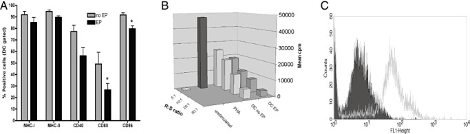
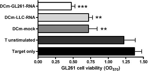
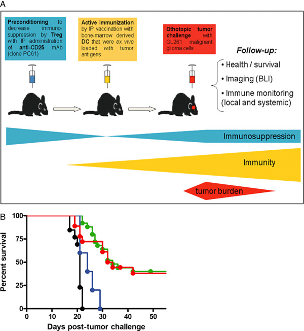
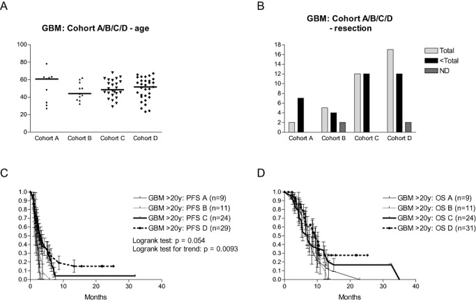
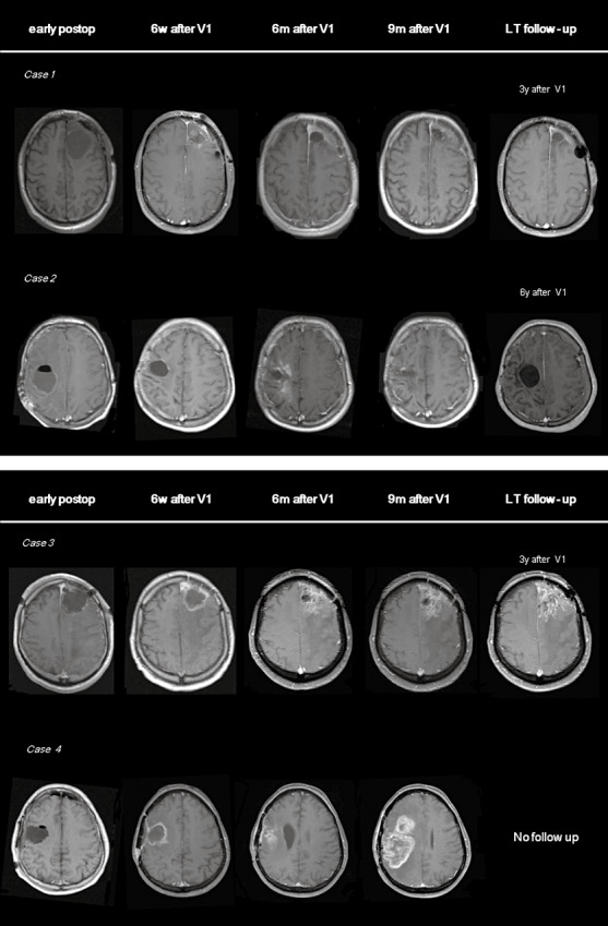
The prognosis of patients with malignant glioma is poor in spite of multimodal treatment approaches consisting of neurosurgery, radiochemotherapy and maintenance chemotherapy. Among innovative treatment strategies like targeted therapy, antiangiogenesis and gene therapy approaches, immunotherapy emerges as a meaningful and feasible treatment approach for inducing long-term survival in at least a subpopulation of these patients. Setting up immunotherapy for an inherent immunosuppressive tumor located in an immune-privileged environment requires integration of a lot of scientific input and knowledge of both tumor immunology and neuro-oncology. The field of immunotherapy is moving into the direction of active specific immunotherapy using autologous dendritic cells (DCs) as vehicle for immunization. In the translational research program of the authors, the whole cascade from bench to bed to bench of active specific immunotherapy for malignant glioma is covered, including proof of principle experiments to demonstrate immunogenicity of patient-derived mature DCs loaded with autologous tumor lysate, preclinical in vivo experiments in a murine orthotopic glioma model, early phase I/II clinical trials for relapsing patients, a phase II trial for patients with newly diagnosed glioblastoma (GBM) for whom immunotherapy is integrated in the current multimodal treatment, and laboratory analyses of patient samples. The strategies and results of this program are discussed in the light of the internationally available scientific literature in this fast-moving field of basic science and translational clinical research.


Figures

Figure 1
Figure 1
Electroporation of DCs with RNA‐molecules is feasible without loss of cell function in vitro. A. Immature DCs were left untouched (no EP) or electroporated (EP) and immediately afterwards matured with 1 µg/mL (Escherichia coli) lipopolysaccharide for 24 hours. Mature DCs were analyzed by flow cytometry for surface marker expression. EP significantly lowered the expression of CD80 and CD86 on DCs (overall analysis of variance P < 0.001, *P < 0.05). B. The capacity to stimulate allogeneic cells was assessed in a mixed lymphocyte reaction in 96 well format with total 2 × 105 splenocytes from naive BALB/C mice as responder (R) cells and DCs from C57BL/6 mice as stimulator (S) cells. Unstimulated cells and phytohemagglutinin (PHA) were used as negative and positive controls, respectively. Responder to stimulator ratios (R : S) ranged from 5:1 to 50:1. After 4 days of culture, thymidine incorporation was measured by pulsing cultures for 18 hours with 1 µCi [3H] thymidine per well. Mean counts per minute (cpm) are depicted. C. One million DCs were electroporated with 1 µg mRNA encoding enhanced green fluorescent protein (eGFP) and eGFP expression (grey curve in FL1‐Height histogram) was assessed by flow cytometry 24 hours after electroporation. The filled curve represents nonelectroporated DCs.
Figure 2
Figure 2
RNA‐loaded DCs induce a T cell‐mediated antitumor immune response in vitro. T cells pooled from spleen and lymph nodes of a naive C57BL/6 mouse were stimulated ex vivo (two stimulation rounds with DC : T cell ratio of 1:10 with addition of 20 U/mL of IL‐2) with either mock‐loaded DCm (DCm‐mock), DCm electroporated with total RNA extracted from Lewis lung carcinoma cells (DCm‐LLC‐RNA) or DCm electroporated with total RNA extracted from GL261 glioma cells (DCm‐GL261‐RNA). Unstimulated T cells were used as the control. Stimulated T cells were then used as effector cells and coincubated with GL261 tumor cells in an effector to target ratio of 10:1. Tumor cell viability was assessed by measuring the metabolization of 3‐(4,5‐dimethylthiazol‐2‐yl)‐2,5‐diphenyltetrazolium bromide to formazan (measured at 570 nm) by living cells after 2 days of co‐culture. Pooled results (n = 3) are depicted as optical density values at 570 nm (OD570) and compared to GL261 target cells without effector cells. Overall analysis of variance P < 0.001; **P < 0.01, ***P < 0.001.
Figure 3
Figure 3
Immunotherapy in the GL261 experimental orthotopic glioma mouse model. A. Overview of the prophylactic DC vaccination model. Adult C57BL/6 mice are orthotopically challenged with 5 × 105 syngeneic GL261 malignant glioma cells injected 1.5 mm posterior and 1 mm lateral from the bregma at a depth of 3 mm under stereotactic guidance. Read‐out consists of monitoring general health parameters (weight and neurological symptoms), overall survival analysis, in vivo bioluminescence (BLI) requiring GL261 cells expressing luciferase in white mutant C57BL/6.Cg.Tyrc‐2J/J mice, and detailed immune monitoring both systemically (in blood, spleen and tumor‐draining cervical lymph nodes) and locally within the brain. Immunotherapy in our preventive treatment setting consists of either active immunization, decreasing regulatory T cell (Treg)‐mediated immune suppression, or combined treatment. Active immunization is performed through intraperitoneal (IP) vaccination on day 14 and day 7 before tumor challenge with one million mature bone‐marrow derived CD11c+ DCs that were loaded ex vivo with tumor antigens (in the form of total RNA). In some experiments, animals were preconditioned with a single intraperitoneal injection (250 µg) of the depleting anti‐CD25 monoclonal antibody (clone PC61). This in vivo depletion of CD25‐expressing cells was performed 1 week before the first vaccination with DC (or 3 weeks before tumor challenge if no DC vaccination was administered). B. Pooled survival data represented as Kaplan–Meier graph. Overall log rank P < 0.001. Survival curves of animals vaccinated with DCm‐GL261‐RNA (green curve, n = 27), DCm‐GL261‐L (DC loaded with GL261 lysate) (red curve, n = 19), DCm‐splenocyte‐RNA (blue curve, n = 5) and untreated animals (black curve, n = 13) are depicted.
Figure 4
Figure 4
HGG‐IMMUNO‐2003 protocol: DC vaccination for adults with relapsed GBM. Patients with relapsed GBM were reoperated (removal of as much tumor tissue as feasible) and received subsequent immunotherapy. Patients were divided among four consecutive cohorts. A. The age (expressed in years on the y‐axis) for the four cohorts is shown, and median age is indicated. B. The number of patients with total resection or less than total resection of relapsed GBM is shown for each cohort (ND = not documented). C. Progression‐free survival (PFS) for the four cohorts of patients. D. Overall survival (OS) for the four cohorts of patients.
Figure 5
Figure 5
MRI findings during and after DC vaccination for relapsed GBM. Transverse T1‐weighted MR images after contrast administration, obtained 1 day after debulking surgery for relapsed GBM (early postoperative period), 6 weeks after the first vaccine (6 w after V1), 6 months after the first vaccine (6 m after V1), 9 months after the first vaccine (9 m after V1) and after long‐term follow‐up (LT follow‐up), 3 to 6 years after the first vaccine according to the case (3–6 years after V1). Case 1 is a patient with a resection in the left frontal lobe. MR imaging showed discrete contrast enhancement around the resection cavity 6 weeks after administration of the first vaccine. No contrast enhancement was documented in follow‐up imaging. Case 2 is a patient after surgery in the right frontal lobe. Transient contrast enhancement was seen at 6 and 9 months after the start of the vaccination therapy. Follow‐up MR scans showed subsequent resolution of the contrast enhancement. Case 3 shows follow‐up MR images in a patient with surgery in the left frontal lobe. Over consecutive time points, the resection cavity collapsed and a comparable area of contrast enhancement has remained stable for 3 years. Case 4 is a patient with a resection in the right frontoparietal region. Subsequent MRI shows an increasing region of contrast enhancement, suggestive of tumor relapse/progression.

References

    1. Aarntzen EH, Figdor CG, Adema GJ, Punt CJ, De Vries IJ (2008) Dendritic cell vaccination and immune monitoring. Cancer Immunol Immunother 57:1559–1568. - PMC - PubMed
    1. Akasaki Y, Black KL, Yu JS (2005) Dendritic cell‐based immunotherapy for malignant gliomas. Expert Rev Neurother 5:497–508. - PubMed
    1. Akasaki Y, Black KL, Yu JS (2005) T cell immunity in patients with malignant glioma: recent progress in dendritic cell‐based immunotherapeutic approaches. Front Biosci 10:2908–2921. - PubMed
    1. Akasaki Y, Liu G, Chung NH, Ehtesham M, Black KL, Yu JS (2004) Induction of a CD4+ T regulatory type 1 response by cyclooxygenase‐2‐overexpressing glioma. J Immunol 173:4352–4359. - PubMed
    1. Aoki H, Mizuno M, Natsume A, Tsugawa T, Tsujimura K, Takahashi T, Yoshida J (2001) Dendritic cells pulsed with tumor extract‐cationic liposome complex increase the induction of cytotoxic T lymphocytes in mouse brain tumor. Cancer Immunol Immunother 50:463–468. - PMC - PubMed

Publication types

Substances