. 2010 Jan;1797(1):71-80.
doi: 10.1016/j.bbabio.2009.08.002. Epub 2009 Sep 8.

Energetic performance is improved by specific activation of K+ fluxes through K(Ca) channels in heart mitochondria

Affiliations

Energetic performance is improved by specific activation of K+ fluxes through K(Ca) channels in heart mitochondria

Miguel A Aon et al. Biochim Biophys Acta. 2010 Jan.

Abstract

Mitochondrial volume regulation depends on K+ movement across the inner membrane and a mitochondrial Ca2+-dependent K+ channel (mitoK(Ca)) reportedly contributes to mitochondrial K+ uniporter activity. Here we utilize a novel K(Ca) channel activator, NS11021, to examine the role of mitoK(Ca) in regulating mitochondrial function by measuring K+ flux, membrane potential (DeltaPsi(m)), light scattering, and respiration in guinea pig heart mitochondria. K+ uptake and the influence of anions were assessed in mitochondria loaded with the K+ sensor PBFI by adding either the chloride (KCl), acetate (KAc), or phosphate (KH2PO4) salts of K+ to energized mitochondria in a sucrose-based medium. K+ fluxes saturated at approximately 10 mM for each salt, attaining maximal rates of 172+/-17, 54+/-2.4, and 33+/-3.8 nmol K+/min/mg in KCl, KAc, or KH2PO4, respectively. NS11021 (50 nM) increased the maximal K+ uptake rate by 2.5-fold in the presence of KH2PO4 or KAc and increased mitochondrial volume, with little effect on DeltaPsi(m). In KCl, NS11021 increased K+ uptake by only 30% and did not increase volume. The effects of NS11021 on K+ uptake were inhibited by the K(Ca) toxins charybdotoxin (200 nM) or paxilline (1 microM). Fifty nanomolar of NS11021 increased the mitochondrial respiratory control ratio (RCR) in KH2PO4, but not in KCl; however, above 1 microM, NS11021 decreased RCR and depolarized DeltaPsi(m). A control compound lacking K(Ca) activator properties did not increase K+ uptake or volume but had similar nonspecific (toxin-insensitive) effects at high concentrations. The results indicate that activating K+ flux through mitoK(Ca) mediates a beneficial effect on energetics that depends on mitochondrial swelling with maintained DeltaPsi(m).


Figures
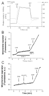
Figure 1
Figure 1
Simultaneous monitoring of mitochondrial volume, ΔΨm, and NAD(P)H, and respiration during state 4 and the state 4state 3 transition in isolated guinea-pig heart mitochondria. A) Freshly isolated mitochondria from guinea-pig heart were resuspended (~100 μg mitochondrial protein) in the cuvette of a spectrofluorometer containing 2ml isosmotic sucrose-based assay medium in the presence of 100nM TMRM with constant stirring at 37°C. At the indicated times, 5mM glutamate-Na+/malate-Na+ (first arrow), or 1mM ADP (second arrow), were added. B, C) Mitochondrial respiration was assayed in a respirometer as described in the methods. An increase in signal represents a decrease in oxygen in the chamber. The uncoupler DNP (50μM) was added to determine maximal uncoupled respiration. In panel C, a representative trace is depicted corresponding to mitochondria preincubated with 50nM NS11021, energized with G/M, and subjected to a pulse of 20mM KH2PO4. Notice the decrease in state 4 respiration due to activation of K+ fluxes and swelling (see Figs. 3B and 4). In panel A, it can be clearly seen that on substrate addition, mitochondria respond with low-amplitude swelling (gray trace), ΔΨm polarization by ~40mV (dashed trace) and a 2-fold NAD(P)H pool reduction black trace) whereas ADP addition has the opposite effect, i.e. a volume decrease, ΔΨm depolarization, and NAD(P)H oxidation. Similar results were obtained in isosmotic 137mM KCl-based medium. Key to abbreviations: G/M, glutamate-Na+/malate-Na+; DNP, dinitrophenol.
Figure 2
Figure 2
Simultaneous monitoring of K+ fluxes, volume, and ΔΨm, following pulses of different K+ salts in isolated mitochondria. Freshly isolated mitochondria from guinea-pig heart, resuspended and assayed as described in the legend of Figure 1, were energized with 5mM G/M (both Na+ neutralized) in isosmotic sucrose-based medium. Under these state 4-respiration conditions, different concentrations of potassium chloride, KCl (A–C), potassium acetate, KAc (D–F), or potassium phosphate KH2PO4 (G–I) were added and the normalized PBFI fluorescence ratio 340/380 (A, D, G), 90o light scattering (B, E, H), and ΔΨm (C, F, I), were simultaneously monitored. The results obtained from two independent experiments are shown. Arrows point to the time of the K+ salt addition.
Figure 3
Figure 3
Kinetics of K+ uptake and its activation by NS11021 in isolated mitochondria from guinea-pig heart. A) The initial rates of K+ uptake (nmol K+/min/mg) after pulses of KCl, KAc, or KH2PO4, at different concentrations, were quantified from traces like those shown in Figure 2 (panels A, D, G). Each point in the plot is the result obtained from three independent mitochondrial isolations. The kinetic parameters K0.5 and Vmax were obtained after non-linear regression analysis according to a Michaelis-Menten type of expression (V = Vmax (S)/K0.5 + (S)) performed in MicroCal Origin. The PBFI ratio 340/380 was related to K+ concentration as described in the methods. B) The initial rates of K+ uptake after addition of 10mM KCl, KAc, or KH2PO4, to state 4 mitochondria in the absence (control) or the presence of 10nM, 50nM, 100nM, 200nM, or 1μM of NS11021. The PBFI ratio, 90° light scattering, and ΔΨm, were monitored simultaneously. K+ fluxes normalized to the control (absence of NS11021) for each K+ salt are shown (two independent experiments).
Figure 4
Figure 4
Charybdotoxin and paxilline sensitivity of K+ uptake activation and mitochondrial volume increase elicited by NS11021. The effects of 200nM ChTx (A, B) or 1μM paxilline (C, D) on K+ uptake (A, C) and mitochondrial swelling (B, D) in the presence of 50nM NS11021 following the addition of 10mM KH2PO4 are shown. The PBFI ratio and 90° light scattering were monitored simultaneously. Arrows point to the time of the K+ salt addition. Representative results from two independent experiments with duplicates or triplicates within each are shown.
Figure 5
Figure 5
Effects of NS11021 on mitochondrial respiration. Freshly isolated mitochondria from guinea-pig heart were assayed for state 4 and state 3 mitochondrial respiration under the same conditions described in the legends of Figures 1 and 2. The RCR (state3/state4) was calculated without (control) or with preincubation of 50nM or 1μM NS11021 and 10 or 20mM KH2PO4 (A) or 10 or 20mM KCl (C) without (A,C) or with (B, D) 200nM ChTx. In panel A, the box plots depict the median (line), 25th and 75th percentile (box) and the minimum and maximum value (bars) whereas only the median with maximal and minimal values are represented in all other panels. Panel A: Control (n= 8, 4 experiments); NS50nM (n= 8; 4 experiments); NS1μM (n=8; 4 experiments). Panel B: Control (n=4; 2 experiments); NS50nM (n= 4; two experiments); NS1μM (n=4; 2 experiments). Panel C: Control (n=4; 2 experiments); NS50nM (n= 4; 2 experiments); NS1μM (n= 4; 2 experiments). Panel D: Control (n= 4; 2 experiments); NS50nM (n= 4; 2 experiments); NS1μM (n= 4; 2 experiments).
Figure 6
Figure 6
Comparative effects of NS11021 and valinomycin before or after addition of K+ salt. Freshly isolated mitochondria from guinea-pig heart were assayed as described in the legend of Figure 2 without or with NS11021 (A) or valinomycin (B–D) preincubation within the 25pM to 1nM range (shown are 50pM, 200pM, 500pM, 1nM Val). Mitochondrial ΔΨm, K+ flux and volume were monitored as described in the legend of Figure 2. Arrows point to the time of 10mM KH2PO4 addition.
Figure 7
Figure 7
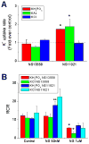
Comparative effects of the KCa activator NS11021 and its inactive congener NS13558 on K+ fluxes and mitochondrial respiration. Freshly isolated mitochondria from guinea-pig heart were assayed under similar conditions as described in the legends of Figures 3 and 5. A) The initial rates of K+ uptake after addition of 10mM KCl, KAc, or KH2PO4, to state 4 mitochondria in the absence (control) or the presence of 50nM or 1μM of NS13558 or NS11021. The PBFI ratio, 90° light scattering, and ΔΨm, were monitored simultaneously in duplicates. Shown are the K+ fluxes normalized to the control (absence of NS compounds) for each K+ salt whereas the other variables are described in Fig. S2 of the Supplement. B) Mitochondria were assayed for state 4 and state 3 respiration in triplicates, and the RCR (state3/state4) calculated without (control) or with preincubation of 50nM or 1μM of NS13558 or NS11021, and 10 or 20mM KH2PO4 or 10 or 20mM KCl.
Figure 8
Figure 8
NS11021 actions on mitochondrial energetics. NS11021 activation of KCa channel-dependent K+ influx (charybdotoxin- and paxilline-inhibitable), increases matrix volume in mitochondria energized with complex I substrates glutamate/malate (G/M) in the absence of ADP (state 4, low respiration, high ΔΨm and NADH). K+ influx and mitochondrial volume are quickly balanced by the K+/H+ antiporter, which expels the cation from the matrix. The specific activation of K+ flux through the KCa channel by NS11021 occurs in the nanomolar concentration range, whereas nonspecific ion leak occurs in the micromolar range. The inactive form NS13558 did not evoke KCa specific actions, but retained the nonspecific effects at micromolar levels. The scheme also highlights that the degree of K+ influx and the increase in volume is dependent upon the anion permeability (H2PO4 and Ac > Cl), and that the valinomycin-activated effects differ from those of the KCa channel. Key to symbols: KCa, Ca2+-dependent K+ channel (mitoKCa); K+/H+, K+/H+ antiporter; ETC, electron transport chain; ChTx, charybdotoxin; Pax, paxilline; IM, inner membrane; OM, outer membrane.

References

    1. Garlid KD, Dos Santos P, Xie ZJ, Costa AD, Paucek P. Mitochondrial potassium transport: the role of the mitochondrial ATP-sensitive K(+) channel in cardiac function and cardioprotection. Biochimica et biophysica acta. 2003;1606:1–21. - PubMed
    1. O’Rourke B. Evidence for mitochondrial K+ channels and their role in cardioprotection. Circ Res. 2004;94:420–432. - PMC - PubMed
    1. Bednarczyk P, Barker GD, Halestrap AP. Determination of the rate of K(+) movement through potassium channels in isolated rat heart and liver mitochondria. Biochimica et biophysica acta. 2008;1777:540–548. - PubMed
    1. Kaasik A, Safiulina D, Zharkovsky A, Veksler V. Regulation of mitochondrial matrix volume. Am J Physiol Cell Physiol. 2007;292:C157–163. - PubMed
    1. Brierley GP. The uptake and extrusion of monovalent cations by isolated heart mitochondria. Mol Cell Biochem. 1976;10:41–63. - PubMed

Publication types