. 2009 Nov-Dec;130(11-12):713-21.
doi: 10.1016/j.mad.2009.08.007.

Limited expansion of virus-specific CD8 T cells in the aged environment

Affiliations

Limited expansion of virus-specific CD8 T cells in the aged environment

Jiu Jiang et al. Mech Ageing Dev. 2009 Nov-Dec.

Abstract

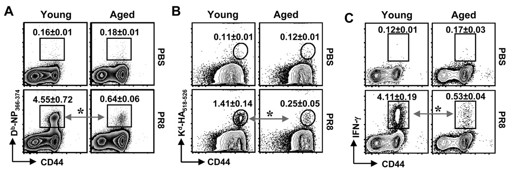
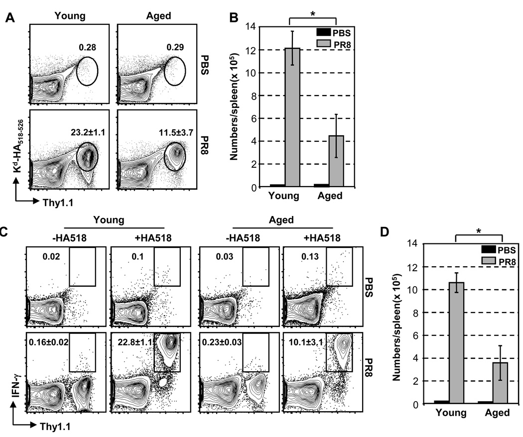
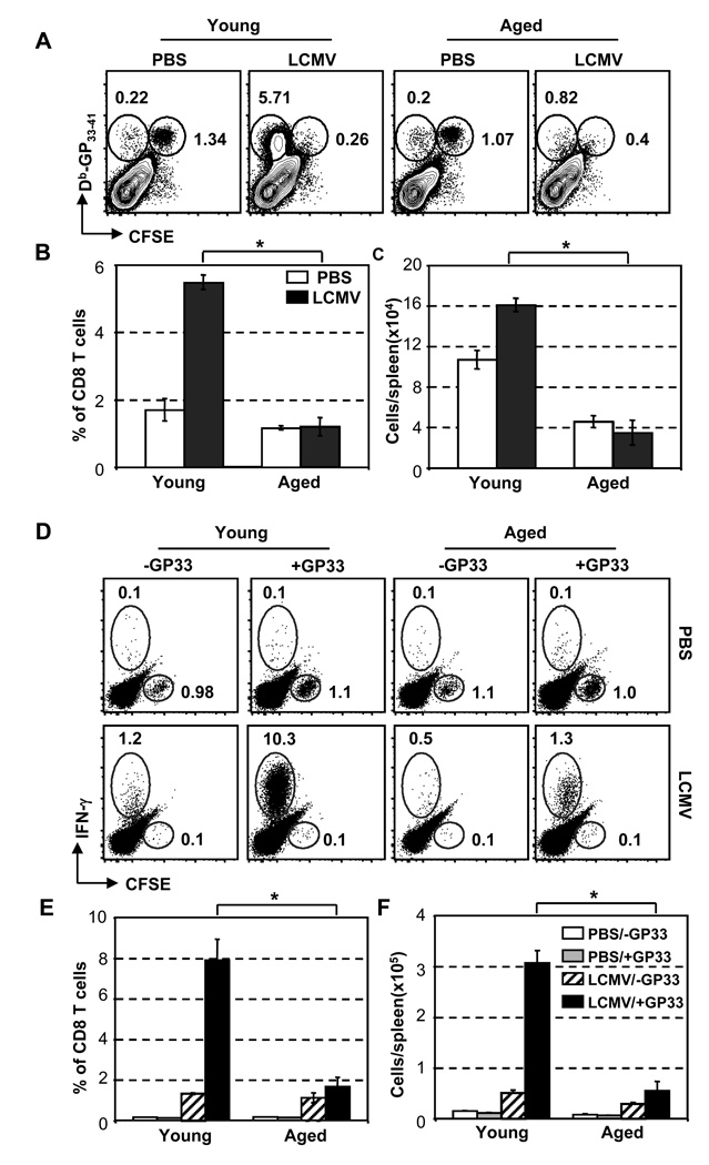
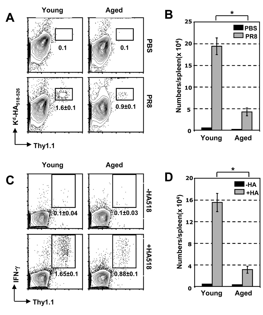
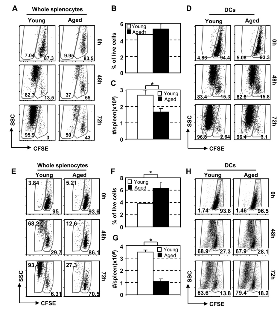
The mechanisms responsible for the diminished immune response seen with aging are unclear. In this study, we investigate the contributions of alterations in the lymphoid microenvironment to this decrease. Using adoptive transfer of virus-specific transgenic CD8 T cells, we demonstrate that the aged environment inhibits the clonal expansion of specific CD8 T cells from young mice during virus infection. Transferred specific CD8 T cells from young mice demonstrated a response reflecting the CD8 T cell response of the intact aged host: the CD8 T cells expand more slowly and have a decreased maximal expansion in an aged compared to a young environment. While isolated DCs (MHC II(+) CD11c(+)) of aged mice maintain their ability to support CD8 T cell Ag-specific expansion in vitro, splenocytes demonstrated an age-associated decrease in this ability. Since the percentages of various populations of DCs in splenocytes demonstrate no significant alteration with age, this diminished APC activity of splenocytes of aged mice may reflect inhibitory activity of other cell populations. The results of this study demonstrate that elements of the aged environment play an important role in the alteration of T cell response to virus infection in the aged.


Figures

Fig. 1
Fig. 1
Ag-specific CD8 T cell response to primary influenza infection is decreased in aged mice. Young and aged B6 (A & C), and BALB/c (B) mice were infected i.v. with 300 HAU PR8. On Day 7 post infection, splenocytes were isolated, stained with CD8, CD44 and anti-IFN-γ antibodies, as well as Db-NP366–374, or Kd-HA tetramers, and assessed by flow cytometry. Fig. A and B: Tetramer+/CD44high cells are presented as percent of total CD8 T cells. Fig. C: IFN-γ production as percentage of cells that are CD8+IFN-γ+ of total CD8+ cells. Each plot shows CD8+ cells from one representative mouse with numbers indicating mean ± SD of 3–4 mice. * p < 0.05. Data are representative of three independent experiments with similar results.
Fig. 2
Fig. 2
Limited expansion of higher numbers of Tg CD8 T cells in response to influenza virus infection in aged BALB/c mice. 2–4 × 10⁶ splenocytes of Clone-4 TCR-Tg mice (Thy1.1+) were adoptively transferred into congenic young and aged BALB/c mice (Thy1.2+). Recipient mice were infected i.v. with 300 HAU PR8. On Day 3 post-infection, splenocytes were isolated, stained with antibodies to CD8, CD44 and IFN-γ, as well as Kd-HA tetramer. Donor CD8 T cells were identified by HA+/Thy1.1+ staining after gating on total CD8 T cells. (A) Percentages of HA-specific donor CD8 T cell of total CD8+ cells. (B) Absolute numbers of HA518–526+/Thy1.1+ cells. (C) Percentage of Thy1.1+IFN-γ+ gated on CD8+ cells. (D) Absolute numbers of Thy1.1+IFN-γ+ cells. Each plot is representative of one mouse with numbers reflecting ± SD of 3–4 mice. * p < 0.05. These results are representative of four independent experiments with similar results.
Fig. 3
Fig. 3
Limited expansion of Db-GP33–41 Tg CD8 T cells in aged B6 mice after infection with LCMV. 2–4 × 10⁶ CFSE-labeled splenocytes from GP33–41-specific P14 T B6 mice were adoptively transferred into young and aged B6 mice. Recipient mice were infected i.p. with 2 ×10⁶ PFU LCMV Arm. On Day 3 post-infection, proliferation and expansion of transferred P14 cells in the spleen were examined by CFSE, antibody and Db-GP33–41 tetramer staining. (Fig. A & B) Percentages of GP33–41+ specific CD8 T cells of total CD8+ cells; (C) Absolute numbers of GP33–41+ CD8 T cells (gated on CD8+ cells); (Fig. D & E) Percentages of IFN-γ+ CD8 T cells, gated on CD8+ cells; (F) Absolute numbers of CD8+IFN-γ+ cells. Each plot is representative of one mouse from 3–4 mice per group. Error bars represent ± SD. * p < 0.05. Similar results were obtained in three separate experiments.
Fig. 4
Fig. 4
Limited expansion of lower numbers of HA518–526 Tg CD8 T cells in response to influenza virus infection in aged BALB/c mice. 5×10³ splenocytes of Clone-4 cells (Thy1.1+) were adoptively transferred into young and aged BALB/c mice (Thy1.2+). The recipients were infected i.v. with 300 HAU PR8. On Day 4 after infection, expansion of donor CD8 T cells in the spleens was identified by HA+Thy1.1+ staining after gating on total CD8 T cells, and the function of the specific CD8 T cells were examined by intracellular IFN-γ staining after in vitro stimulation with HA518–526 peptide for 5 h. (A) Percentages of HA-specific donor CD8 T cell of total CD8+ cells. (B) Absolute numbers of HA518–526+Thy1.1+ cells. (C) Percentage of Thy1.1+IFN-γ+ gated on CD8+ cells. (D) Absolute numbers of Thy1.1+IFN-γ+ cells. Each plot is representative of one mouse with numbers reflecting ± SD of 3–4 mice. * p < 0.05. Similar results were obtained in three independent experiments.
Fig. 5
Fig. 5
The ability of splenocytes of aged mice to support expansion of specific CD8 T cells is lower compared to splenocytes of young mice. (A–D) Clone-4 Tg mice and BALB/c mice: (A) 1 × 10⁵ CFSE-labeled purified Clone-4 CD8 T cells were cultured with HA518–526 and 5 × 10⁵ splenocytes from young or aged BALB/c mice. Proliferation and expansion of the TCR-Tg T cells (gated on Thy1.1+CD8+); (B) Percentages, and (C) Numbers of CD11C+/MHC I-Ad+ cells in the spleen of BALB/c mice; (D) 1 × 10⁵ CFSE-labeled Clone-4 CD8 T cells (Thy1.1+) were cultured with 1× 10⁴ enriched DCs from young or aged BALB/c mice (Thy1.2+). Proliferation and expansion of the TCR-Tg T cells. (E–H): P14 Tg mice and B6 mice: (E) 1 × 10⁵ CFSE-labeled purified P14 CD8 T cells were cultured with 5 × 10⁵ splenocytes from young or aged B6 mice. Proliferation and expansion of the P14 cells (gated on CD8+GP33–41+); (F) Percentages, and (G) Numbers of CD11C+/MHC I-Ab+ cells in the spleen of B6 mice; (H) 1 × 10⁵ CFSE labeled P14 CD8 T cells were incubated with 1× 10⁴ enriched DCs from young or aged B6 mice. Proliferation and expansion of P14 cells. N = 3 in each group. Error bars represent ± SD. * p < 0.05. Data are representative of three independent experiments with similar results.

References

    1. Appay V, Rowland-Jones SL. The assessment of antigen-specific CD8+ T cells through the combination of MHC class I tetramer and intracellular staining. J. Immunol. Methods. 2002;268:9–19. - PubMed
    1. Ashton-Rickardt PG, Bandeira A, Delaney JR, Van Kaer L, Pircher HP, Zinkernagel RM, Tonegawa S. Evidence for a differential avidity model of T cell selection in the thymus. Cell. 1994;76:651–663. - PubMed
    1. Badovinac VP, Haring JS, Harty JT. Initial T cell receptor transgenic cell precursor frequency dictates critical aspects of the CD8(+) T cell response to infection. Immunity. 2007;26:827–841. - PMC - PubMed
    1. Beharka AA, Wu D, Han SN, Meydani SN. Macrophage prostaglandin production contributes to the age-associated decrease in T cell function which is reversed by the dietary antioxidant vitamin E. Mech. Ageing Dev. 1997;93:59–77. - PubMed
    1. Boulter JM, Schmitz N, Sewell AK, Godkin AJ, Bachmann MF, Gallimore AM. Potent T cell agonism mediated by a very rapid TCR/pMHC interaction. Eur. J. Immunol. 2007;37:798–806. - PMC - PubMed

Publication types