

Nuclear dynamics of influenza A virus ribonucleoproteins revealed by live-cell imaging studies
- PMID: 19744689
- PMCID: PMC2771073
- DOI: 10.1016/j.virol.2009.08.015
Nuclear dynamics of influenza A virus ribonucleoproteins revealed by live-cell imaging studies
Abstract
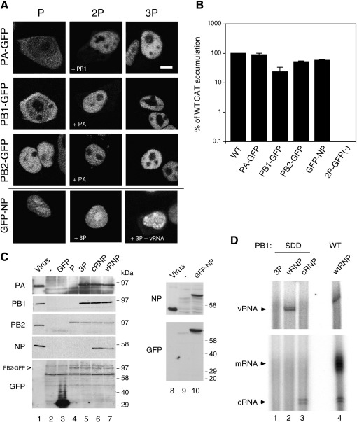
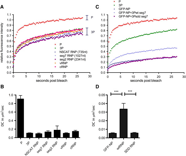
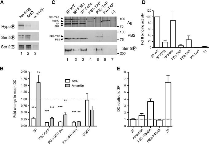
The negative sense RNA genome of influenza A virus is transcribed and replicated in the nuclei of infected cells by the viral RNA polymerase. Only four viral polypeptides are required but multiple cellular components are potentially involved. We used fluorescence recovery after photobleaching (FRAP) to characterise the dynamics of GFP-tagged viral ribonucleoprotein (RNP) components in living cells. The nucleoprotein (NP) displayed very slow mobility that significantly increased on formation of transcriptionally active RNPs. Conversely, single or dimeric polymerase subunits showed fast nuclear dynamics that decreased upon formation of heterotrimers, suggesting increased interaction of the full polymerase complex with a relatively immobile cellular component(s). Treatment with inhibitors of cellular transcription indicated that in part, this reflected an interaction with cellular RNA polymerase II. Analysis of mutated influenza virus polymerase complexes further suggested that this was through an interaction between PB2 and RNA Pol II separate from PB2 cap-binding activity.
Figures


Similar articles
-
Eukaryotic Translation Elongation Factor 1 Delta Inhibits the Nuclear Import of the Nucleoprotein and PA-PB1 Heterodimer of Influenza A Virus.J Virol. 2020 Dec 22;95(2):e01391-20. doi: 10.1128/JVI.01391-20. Print 2020 Dec 22. J Virol. 2020. PMID: 33087462 Free PMC article.
-
Influenza polymerase activity correlates with the strength of interaction between nucleoprotein and PB2 through the host-specific residue K/E627.PLoS One. 2012;7(5):e36415. doi: 10.1371/journal.pone.0036415. Epub 2012 May 3. PLoS One. 2012. PMID: 22570712 Free PMC article.
-
Interaction between influenza A virus nucleoprotein and PB2 cap-binding domain is mediated by RNA.PLoS One. 2020 Sep 28;15(9):e0239899. doi: 10.1371/journal.pone.0239899. eCollection 2020. PLoS One. 2020. PMID: 32986763 Free PMC article.
-
Insight into Influenza: A Virus Cap-Snatching.Viruses. 2018 Nov 16;10(11):641. doi: 10.3390/v10110641. Viruses. 2018. PMID: 30453478 Free PMC article. Review.
-
Structure and assembly of the influenza A virus ribonucleoprotein complex.FEBS Lett. 2013 Apr 17;587(8):1206-14. doi: 10.1016/j.febslet.2013.02.048. Epub 2013 Mar 13. FEBS Lett. 2013. PMID: 23499938 Review.
Cited by
-
Compounds with anti-influenza activity: present and future of strategies for the optimal treatment and management of influenza. Part I: Influenza life-cycle and currently available drugs.J Prev Med Hyg. 2014 Sep;55(3):69-85. J Prev Med Hyg. 2014. PMID: 25902573 Free PMC article. Review.
-
Transport of the influenza virus genome from nucleus to nucleus.Viruses. 2013 Oct 2;5(10):2424-46. doi: 10.3390/v5102424. Viruses. 2013. PMID: 24104053 Free PMC article. Review.
-
Poly-ADP Ribosyl Polymerase 1 (PARP1) Regulates Influenza A Virus Polymerase.Adv Virol. 2019 Mar 19;2019:8512363. doi: 10.1155/2019/8512363. eCollection 2019. Adv Virol. 2019. PMID: 31015836 Free PMC article.
-
Structural basis of an essential interaction between influenza polymerase and Pol II CTD.Nature. 2017 Jan 5;541(7635):117-121. doi: 10.1038/nature20594. Epub 2016 Dec 21. Nature. 2017. PMID: 28002402
-
RNA-Free and Ribonucleoprotein-Associated Influenza Virus Polymerases Directly Bind the Serine-5-Phosphorylated Carboxyl-Terminal Domain of Host RNA Polymerase II.J Virol. 2016 Jun 10;90(13):6014-6021. doi: 10.1128/JVI.00494-16. Print 2016 Jul 1. J Virol. 2016. PMID: 27099314 Free PMC article.
References
Publication types
MeSH terms
Substances
Grants and funding
LinkOut - more resources
Full Text Sources
Other Literature Sources
Miscellaneous
