. 2009 Nov 6;284(45):31122-9.
doi: 10.1074/jbc.M109.001347. Epub 2009 Sep 10.

Human Naa50p (Nat5/San) displays both protein N alpha- and N epsilon-acetyltransferase activity

Affiliations

Human Naa50p (Nat5/San) displays both protein N alpha- and N epsilon-acetyltransferase activity

Rune Evjenth et al. J Biol Chem. .

Abstract

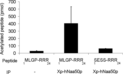
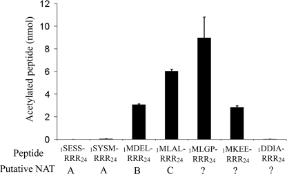
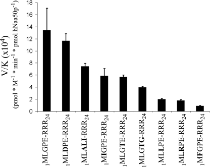
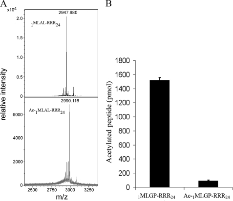
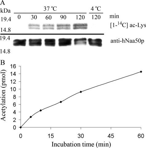
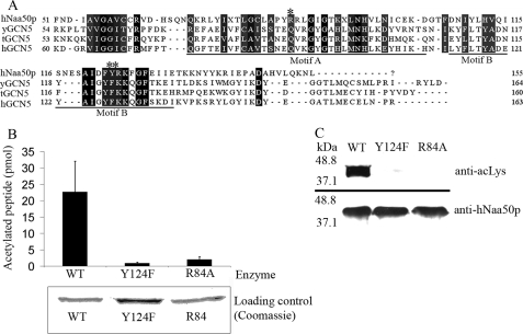
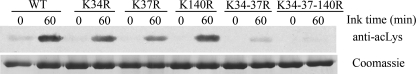
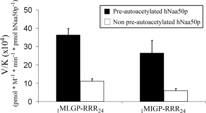
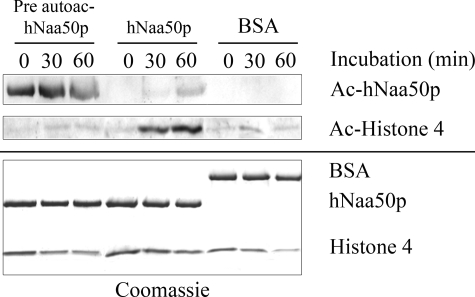
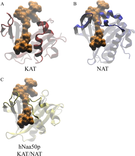
Protein acetylation is a widespread modification that is mediated by site-selective acetyltransferases. KATs (lysine N(epsilon)-acetyltransferases), modify the side chain of specific lysines on histones and other proteins, a central process in regulating gene expression. N(alpha)-terminal acetylation occurs on the ribosome where the alpha amino group of nascent polypeptides is acetylated by NATs (N-terminal acetyltransferase). In yeast, three different NAT complexes were identified NatA, NatB, and NatC. NatA is composed of two main subunits, the catalytic subunit Naa10p (Ard1p) and Naa15p (Nat1p). Naa50p (Nat5) is physically associated with NatA. In man, hNaa50p was shown to have acetyltransferase activity and to be important for chromosome segregation. In this study, we used purified recombinant hNaa50p and multiple oligopeptide substrates to identify and characterize an N(alpha)-acetyltransferase activity of hNaa50p. As the preferred substrate this activity acetylates oligopeptides with N termini Met-Leu-Xxx-Pro. Furthermore, hNaa50p autoacetylates lysines 34, 37, and 140 in vitro, modulating hNaa50p substrate specificity. In addition, histone 4 was detected as a hNaa50p KAT substrate in vitro. Our findings thus provide the first experimental evidence of an enzyme having both KAT and NAT activities.


Figures

FIGURE 1.
FIGURE 1.
In vitro acetyltransferase activity of immunoprecipitated Xpress-hNaa50p. Immunoprecipitated Xpress-hNaa50p from transfected human embryonic kidney 293 cells was incubated with [1-14C]acetyl-CoA and the oligopeptides 1MLGP-RRR24 or 1SESS-RRR24 for 2 h at 37 °C. dH2O used as negative control (−). After incubation, the oligopeptides were isolated with SP-Sepharose beads, washed three times with 0.5 m acetic acid, and analyzed with scintillation counting. Error bars (S.D.) are based on three independent experiments. For details see “Experimental Procedures.”
FIGURE 2.
FIGURE 2.
hNaa50p/hSan acetylation of peptides with the N termini Met-Leu or Met-Asp. Purified GST-hNaa50p was incubated with selected oligopeptides and [1-14C]acetyl-CoA for 2 h at 37 °C. After incubation, the oligopeptides were isolated with SP-Sepharose beads, washed three times with 0.5 m acetic acid, and analyzed with scintillation counting. Activity detected in the negative control (dH2O) was subtracted. The NAT complexes that are expected to perform the acetylation in vivo are given. Error bars (S.D.) are based on three independent experiments. For details see “Experimental Procedures.”
FIGURE 3.
FIGURE 3.
GST-hNaa50p specificity constants (V/K). 70 nm GST-hNaa50p was incubated with selected oligopeptide substrates for 30 min at 37 °C with saturated levels of acetyl-CoA (300 μm). The acetylation kinetics were determined with reverse phase HPLC. V/K is the Vmax/Km (oligopeptides). Error bars indicate the S.D. Experiments were performed in triplicate. For details see “Experimental Procedures.”
FIGURE 4.
FIGURE 4.
Verification of hNaa50p Nα-acetyltransferase activity. A, MALDI-MS spectra (mass range 2.400–3.350) of non-acetylated (upper panel), and acetylated 1MLAL-RRR24 peptide (lower panel) after in vitro acetylation with GST-hNaa50p. Monoisotopic peaks are labeled with their respective m/z ratios. B, purified GST-hNaa50p was incubated for 2 h at 37 °C with 1MLGP-RRR24 peptide and a chemically N-terminal acetylated version of the 1MLGP-RRR24 peptide (Ac-1MLGP-RRR24). After incubation, the oligopeptides were isolated with SP-Sepharose beads, washed three times in 0.5 m acetic acid, and analyzed with scintillation counting. Error bars (S.D.) are based on three independent experiments.
FIGURE 5.
FIGURE 5.
The in vitro autoacetyltransferase activity of purified wild type hNaa50p. A, purified hNaa50p was incubated with [1-14C]acetyl-CoA for the indicated times and analyzed by autoradiography and Western blotting with anti-hNaa50p antibody. Upper panel indicates the increased level of [1-14C]acetyl incorporation as a response to increased incubation time at 37 °C. No acetylation is detectable in the sample incubated for 120 min at 4 °C. Lower panel represents Western blot analyses with anti-hNaa50p showing an equal amount of hNaa50p present in the assay. B, purified GST-hNaa50p (3 μm) was incubated with acetyl-CoA (500 μm with 33% radioactive acetyl-CoA) at 37 °C, and samples were collected at 6 different time points. After isolating the oligopeptides with reverse phase HPLC, the acetylation was quantified by scintillation counting of incorporated radioactivity.
FIGURE 6.
FIGURE 6.
Enzymatic activity of hNaa50p Arg84 to Ala or Tyr124 to Phe mutants. A, alignment of amino acid sequences of hNaa50p with GCN5 from Saccharomyces cerevisiae (yGCN5), Tetrahymena (tGCN5), and human (hGCN5). The alignment indicates that Arg84 (*) and Tyr124 (**) are located in Motif A and Motif B, respectively, of the GCN5 protein fold. B, in vitro acetyltransferase assay comparing the enzyme activity of purified GST-hNat5 WT with GST-hNat5 Arg84 to Ala mutant (R84A) and GST-hNat5 Tyr124 to Phe mutant (Y124F). The 1MLGP-RRR24 oligopeptide (30 μm) was used as substrate. Error bars (S.D.) are based on three independent experiments. Coomassie-stained SDS-PAGE verifying an equal amount of each enzyme. C, purified GST-hNaa50p WT and mutants were incubated for 1 h 37 °C with acetyl-CoA. Western blot analyses using anti-acetylated lysine (anti-acLys) antibody showed a significant difference in the autoacetylation pattern between the WT and mutants. An equal protein amount is shown by Western blotting using anti-hNaa50p.
FIGURE 7.
FIGURE 7.
Comparison of the autoacetylation pattern for GST-hNaa50p WT and different lysine mutants. Upper panel, Western blotting analyses of GST-hNaa50p WT and different lysine to arginine mutants using anti-acetylated lysine (anti-acLys) antibody. 2.4 μm enzymes were incubated for 1 h at 37 °C with 500 μm acetyl-CoA. Lower panel, Coomassie-stained SDS-PAGE presenting protein loading.
FIGURE 8.
FIGURE 8.
The specificity constants (V/K) for two selected in vitro oligopeptide substrates using preautoacetylated and non-preautoacetylated GST-hNaa50p. 10 μm purified GST-hNaa50p was incubated for 5 days at 37 °C with either 300 μm acetyl-CoA (Pre-autoacetylated) or H2O (Non pre-autoacetylated). The Km values for 1MLGP-RRR24 or 1MIGP-RRR24 were then determined with these enzyme variants. 30 nm GST-hNaa50p were incubated with either 1MLGP-RRR24 (20–400 μm) or 1MIGP-RRR24 (50–500 μm) oligopeptide substrate at 37 °C for 30 min with saturated levels of acetyl-CoA (300 μm). The acetylation kinetics were determined with reverse phase HPLC. V/K is the Vmax/Km (oligopeptides). Error bars indicate the S.D. Experiments were performed in triplicates.
FIGURE 9.
FIGURE 9.
GST-hNaa50p KAT activity on histone 4. 10 μm purified GST-hNaa50p was incubated for 5 days at 37 °C with either 300 μm acetyl-CoA (Pre-autoacetylated) or H2O (Non pre-autoacetylated). 2 μm of these enzyme variants were then tested for KAT activity toward Histone 4 protein (2 μm). Equal amounts of bovine serum albumin (BSA) replaced hNaa50p in the negative control. The samples were incubated at 37 °C, and aliquots were collected as indicated. Upper panel, Western blotting analyses of GST-hNaa50ps KAT activity using anti-acetylated lysine (anti-acLys) antibody. Lower panel, Coomassie-stained SDS-PAGE presenting the protein loading.
FIGURE 10.
FIGURE 10.
Structural superimposing of hNaa50p, RimL from S. thyphimurium (NAT), and GNAT enzyme from B. cereus (histone acetyltransferases). A, protein structure of GNAT (B. cereus; PDB code 3BLN) was structurally compared with tGCN5 (Tetrahymena; PDB code 1PU9) that had been co-crystallized with a stretch of the H3 substrate (residue 5–23) shown in orange. B, protein structure of RimL (S. thyphimurium; PDB code 1S7N) with the same H3 substrate stretch present in orange. C, structural comparison of hNaa50p (PDB code 2OB0) with the H3 substrate stretch in orange. All structures are with similar orientations, and parts the protein structures are indicated as partly transparent to clarify the point.

References

    1. Marmorstein R. (2004) Methods Enzymol. 376, 106–119 - PubMed
    1. Kalkhoven E. (2004) Biochem. Pharmacol. 68, 1145–1155 - PubMed
    1. Polevoda B., Brown S., Cardillo T. S., Rigby S., Sherman F. (2008) J. Cell. Biochem. 103, 492–508 - PubMed
    1. Gautschi M., Just S., Mun A., Ross S., Rücknagel P., Dubaquié Y., Ehrenhofer-Murray A., Rospert S. (2003) Mol. Cell. Biol. 23, 7403–7414 - PMC - PubMed
    1. Polevoda B., Sherman F. (2003) J. Mol. Biol. 325, 595–622 - PubMed

Publication types