. 2009 Sep 11;325(5946):1384-7.
doi: 10.1126/science.1176099.

A dimeric structure for archaeal box C/D small ribonucleoproteins

Affiliations

A dimeric structure for archaeal box C/D small ribonucleoproteins

Franziska Bleichert et al. Science. .

Abstract

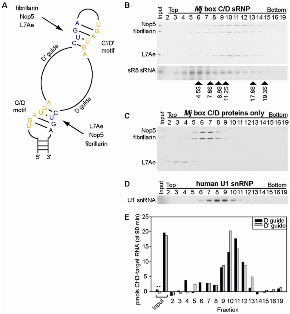
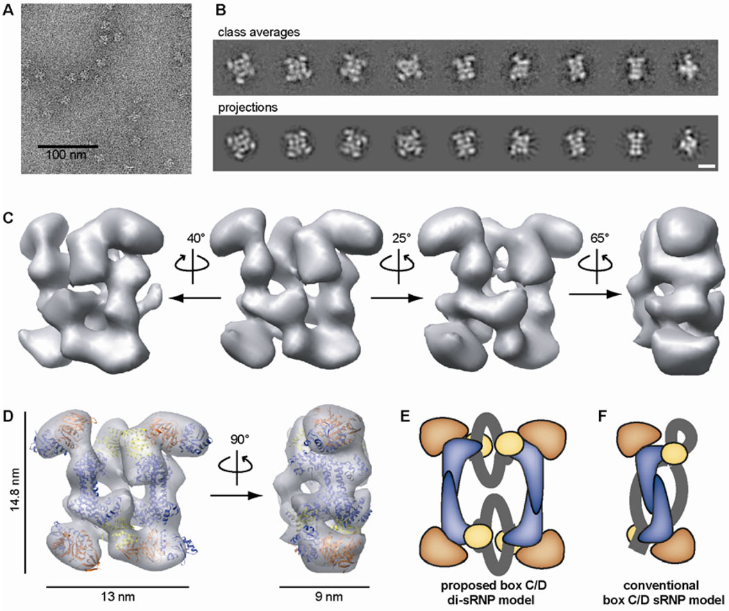
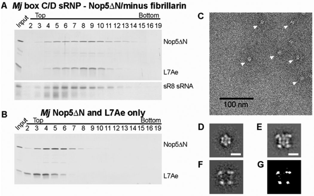
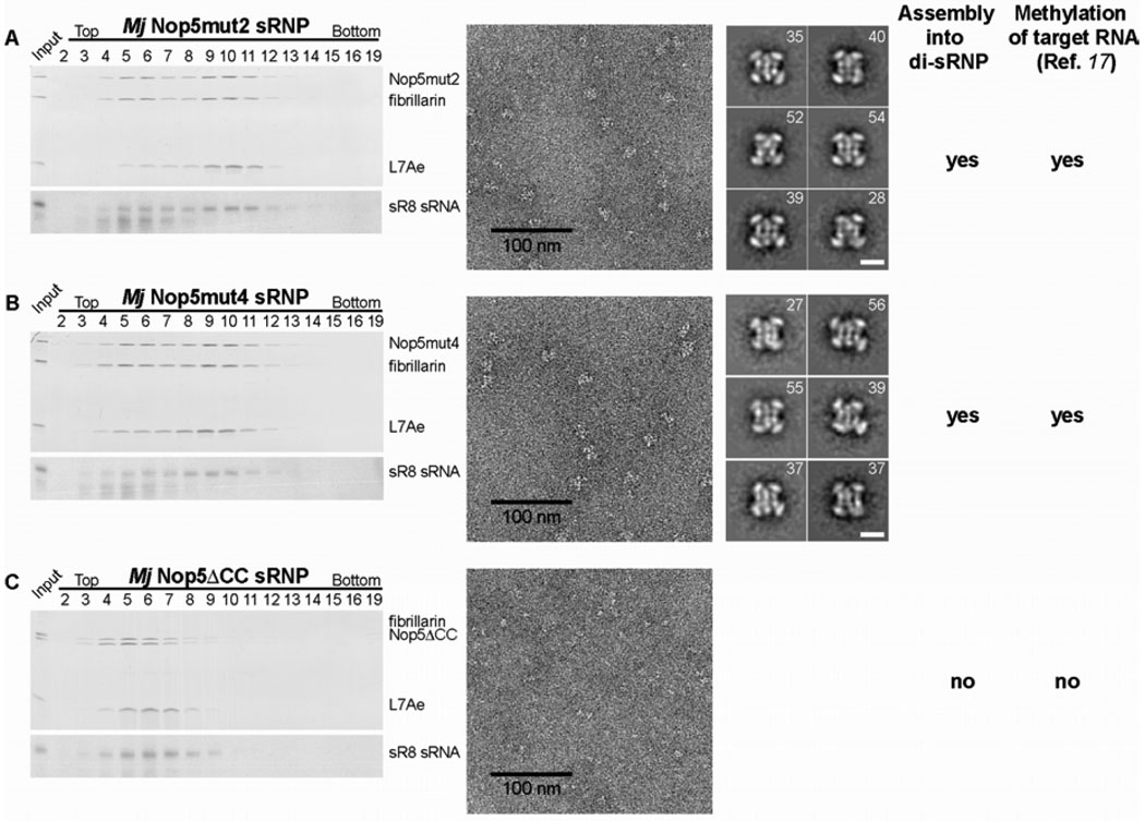
Methylation of ribosomal RNA (rRNA) is required for optimal protein synthesis. Multiple 2'-O-ribose methylations are carried out by box C/D guide ribonucleoproteins [small ribonucleoproteins (sRNPs) and small nucleolar ribonucleoproteins (snoRNPs)], which are conserved from archaea to eukaryotes. Methylation is dictated by base pairing between the specific guide RNA component of the sRNP or snoRNP and the target rRNA. We determined the structure of a reconstituted and catalytically active box C/D sRNP from the archaeon Methanocaldococcus jannaschii by single-particle electron microscopy. We found that archaeal box C/D sRNPs unexpectedly formed a dimeric structure with an alternative organization of their RNA and protein components that challenges the conventional view of their architecture. Mutational analysis demonstrated that this di-sRNP structure was relevant for the enzymatic function of archaeal box C/D sRNPs.


Figures

Fig. 1
Fig. 1
Assembly, purification, and enzymatic activity of the reconstituted Mj box C/D sR8 sRNP. (A) Schematic of an archaeal box C/D sRNA. The conserved sequence motifs, boxes C and C' (yellow) and boxes D and D' (blue), as well as the guide sequences are indicated. Each box C/D and box C'/D' motif is bound by the core box C/D proteins, L7Ae, Nop5, and fibrillarin. (B) Purification of reconstituted sR8 sRNP by glycerol gradient centrifugation. Unpurified sRNP (input) and harvested fractions, as indicated, were analyzed for the presence of the protein components by SDS-PAGE and silver staining (top) and of the sRNA component by Northern blotting (bottom). Arrowheads below the gel indicate peaks of protein markers of corresponding S values run in a parallel gradient. (C) Sedimentation of box C/D sRNP protein components in the absence of the box C/D sRNA in glycerol gradients as carried out in (B). (D) Sedimentation of the human U1 snRNP (MW 240 kDa) in a glycerol gradient run in parallel to (B) using the same experimental conditions. (E) Methylation activity of the Mj box C/D sR8 sRNP in the unpurified material (input) and in the gradient fractions using substrate RNAs complementary to the D guide (black bars) and D' guide (gray bars) sequences of the sR8 sRNA, respectively. As a control for non-specific methylation activity, pre-methylated RNAs were used as substrates (*).
Fig. 2
Fig. 2
Electron microscopy and 3D reconstruction of the archaeal box C/D sR8 sRNP. (A) Electron micrograph of negatively stained sR8 sRNPs from peak fractions after glycerol gradient centrifugation. (B) Experimental class averages and corresponding 2D projections of the reconstructed 3D volume. Scale bar is 10 nm. (see also figs. S3 and S4) (C) Isodensity map of the reconstructed 3D volume of the archaeal box C/D sRNP rotated around the y-axis as indicated. (D) Docking of the crystal structures of Pyrococcus furiosus Nop5-fibrillarin (PDB 2nnw, 16) and Mj L7Ae (1xbi, 31) in the isodensity map. Nop5 - blue, fibrillarin - orange, L7Ae - yellow. The volumes are thresholded to 118% of the MW of the di-sRNP. (E) Proposed di-sRNP model contrasted with (F) the conventional model of archaeal box C/D sRNP architecture. Note that the Nop5-fibrillarin heterotetramer is seen along its 2-fold symmetry axis in the conventional model, whereas the Nop5-fibrillarin heterotetramer in the di-sRNP model is rotated 90° clockwise with respect to the view shown in the conventional model. Colors are as in (D) and the RNA is shown in grey. The orientation of the sRNA ends was purposefully left ambiguous.
Fig. 3
Fig. 3
Localization of fibrillarin and the N-terminal domain of Nop5 in the EM structure. Sedimentation profile of (A) the Nop5ΔN/minus fibrillarin sRNP and (B) L7Ae and Nop5ΔN in the absence of the sR8 sRNA analyzed by glycerol gradient centrifugation. Glycerol gradient centrifugation was performed and analyzed as described in Fig. 1B. (C) Electron micrograph and (D) representative class average of negatively stained Nop5ΔN/minus fibrillarin sRNP particles from peak glycerol gradient fractions. Particles are marked with white arrowheads in (C). See also fig. S11 for full-sized images and additional class averages. (E) Class average of the reconstituted box C/D sRNP particles containing fibrillarin and the N-terminus of Nop5. Additional class averages are provided in fig. S3A. Scale bars are 10 nm in (D) and (E). (F) Difference map between the class averages of the two different RNPs as shown in (D) and (E). (G) Statistically significant region with a p-value of p≤0.001.
Fig. 4
Fig. 4
Formation of the box C/D di-sRNP structure correlates with efficient enzymatic activity. Box C/D sRNPs assembled with Nop5 containing point mutations in the coiled coil domain [mut2 in (A) and mut4 in (B)] or a deletion of the coiled-coil domain [ΔCC in (C)] were analyzed by glycerol gradient centrifugation (as in Fig. 1B) and complexes in peak fractions were visualized by electron microscopy after negative staining. Representative class averages of sRNP particles assembled with Nop5mut2 and Nop5mut4, respectively, are shown. The number of images in each class is indicated in the right upper corner of each class average. The sRNP assembled with Nop5ΔCC was too small for accurate image processing and calculation of meaningful class averages.

References

    1. Decatur WA, Fournier MJ. Trends Biochem Sci. 2002;27:344. - PubMed
    1. Liang XH, Liu Q, Fournier MJ. Mol Cell. 2007;28:965. - PubMed
    1. Kiss-Laszlo Z, Henry Y, Bachellerie JP, Caizergues-Ferrer M, Kiss T. Cell. 1996;85:1077. - PubMed
    1. Omer AD, et al. Science. 2000;288:517. - PubMed
    1. Matera AG, Terns RM, Terns MP. Nat Rev Mol Cell Biol. 2007;8:209. - PubMed

Publication types

MeSH terms