. 2009 Sep 11;325(5946):1402-5.
doi: 10.1126/science.1173474.

A G protein-coupled receptor is essential for Schwann cells to initiate myelination

Affiliations

A G protein-coupled receptor is essential for Schwann cells to initiate myelination

Kelly R Monk et al. Science. .

Abstract

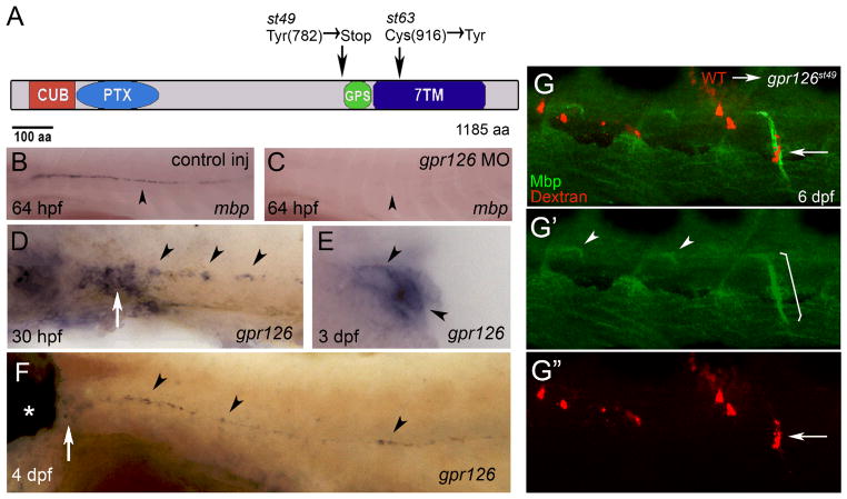
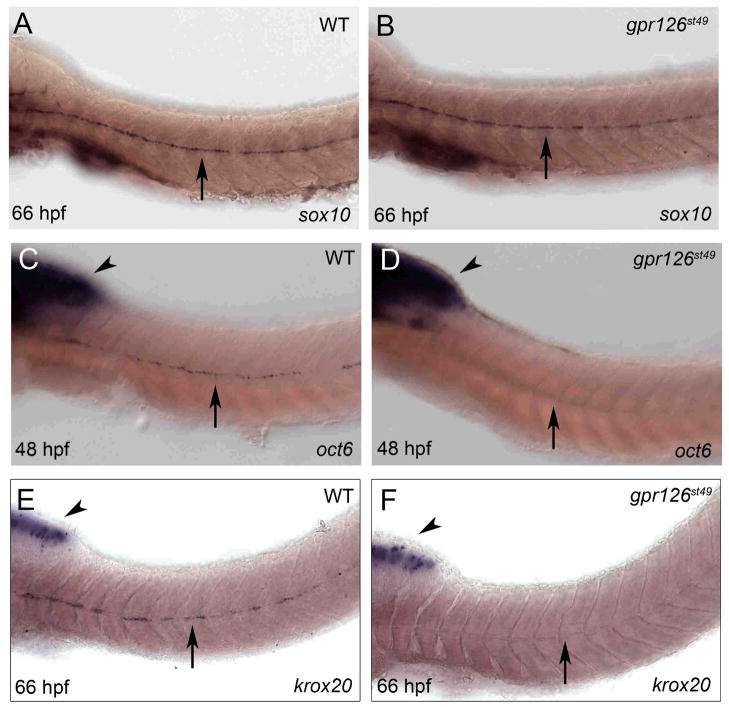
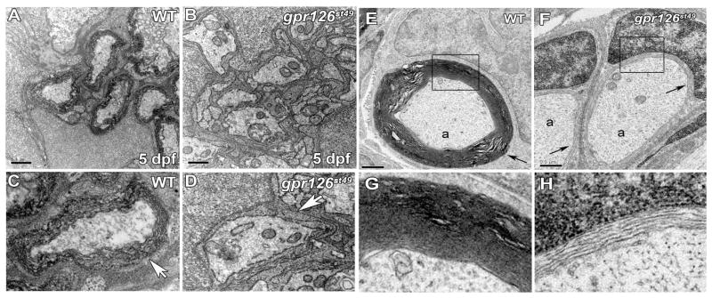
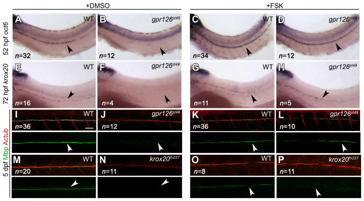
The myelin sheath allows axons to conduct action potentials rapidly in the vertebrate nervous system. Axonal signals activate expression of specific transcription factors, including Oct6 and Krox20, that initiate myelination in Schwann cells. Elevation of cyclic adenosine monophosphate (cAMP) can mimic axonal contact in vitro, but the mechanisms that regulate cAMP levels in vivo are unknown. Using mutational analysis in zebrafish, we found that the G protein-coupled receptor Gpr126 is required autonomously in Schwann cells for myelination. In gpr126 mutants, Schwann cells failed to express oct6 and krox20 and were arrested at the promyelinating stage. Elevation of cAMP in gpr126 mutants, but not krox20 mutants, could restore myelination. We propose that Gpr126 drives the differentiation of promyelinating Schwann cells by elevating cAMP levels, thereby triggering Oct6 expression and myelination.


Figures

Fig. 1
Fig. 1
The st49 and st63 lesions disrupt zebrafish gpr126, which is required autonomously in Schwann cells for Mbp expression. (A) Schematic representation of Gpr126 showing functional domains and the lesions in the st49 and st63 mutations. (B) WT zebrafish embryo injected with control morpholino (MO) showing mbp mRNA in PLLn Schwann cells at 64 hpf (n=13/13). (C) WT zebrafish embryo injected with gpr126 MO showing lack of mbp mRNA in PLLn Schwann cells at 64 hpf (n=19/21). (D–F) Expression of gpr126 in wild-type (WT) larvae examined by whole-mount in situ hybridization. (D) gpr126 mRNA in PLLn Schwann cells (arrowheads) and PLL ganglion (PLLg; arrow) at 30 hpf. (E) Cross-section of a WT larva at 3 dpf showing gpr126 expression in PLLn Schwann cells (arrowheads). (F) gpr126 expression in the ear (*), PLLg (arrow), and in PLLn Schwann cells (arrowheads) at 4 days postfertilization (dpf). For (B–D and F), lateral views are shown, with anterior left and dorsal top. (G) Lateral views at 6 dpf of a chimeric larva generated by transplantation of Texas red dextran-labeled WT cells (red) into a gpr126st49 mutant. WT Schwann cells (arrow in G, G”) express Mbp (green) when associated with mutant motor axons (bracket in G′). In motor nerves without transplanted WT Schwann cells, Mbp expression is only observed at motor nerve exit points as described in Fig. S1 (arrowheads, G′).
Fig. 2
Fig. 2
gpr126 is essential for expression of oct6 and krox20 in Schwann cells. Whole mount in situ hybridization of (A) WT and (B) gpr126st49 mutant zebrafish larvae at 66 hpf showing sox10 mRNA expression in PLLn Schwann cells (arrow). (C) WT zebrafish embryo at 48 hpf showing oct6 expression in the brain (arrowhead) and in PLLn Schwann cells (arrow) as previously described (22). (D) gpr126st49 mutant embryo at 48 hpf showing normal oct6 expression in the brain (arrowhead) but not in PLLn Schwann cells (arrow). (E) WT zebrafish larva at 66 hpf showing krox20 mRNA expression in the brain (arrowhead) and in PLLn Schwann cells (arrow). (F) gpr126st49 mutant larva at 66 hpf showing normal krox20 expression in the brain (arrowhead) but not in PLLn Schwann cells (arrow). For (A–F), anterior is left and dorsal is up. Genotypes were determined by PCR after photography; see Fig. S7 for quantification of these experiments.
Fig. 3
Fig. 3
Schwann cells sort, but do not myelinate axons in gpr126st49 mutants. (A) Transmission electron micrograph showing cross-section through a WT PLLn at 5 dpf. (B) Transmission electron micrograph showing cross-section through a gpr126st49 mutant PLLn at 5 dpf. (C) Magnified view of a WT axon wrapped by several layers of myelin (arrow). (D) Magnified view of a gpr126st49 mutant axon showing that Schwann cell cytoplasm surrounds the axon (arrow), but does not turn more than 1.5 times around the axon. For (A, B), scale bar = 0.5 μm. (E) Transmission electron micrograph showing cross-section through a WT PLLn at 6 months of age. An axon (a) surrounded by compact myelin (arrow) is shown. (F) Transmission electron micrograph showing cross-section through a gpr126st49 mutant PLLn at 6 months of age. Axons (a) surrounded by a few loose wraps of Schwann cell cytoplasm (arrow) are shown. For (E) and (F), scale bar = 0.5 μm. (G) Magnified view of boxed region in (E) showing compact myelin surrounding an axon in WT PLLn. (H) Magnified view of boxed region in (F) showing loose Schwann cell cytoplasm surrounding an axon in gpr126st49 mutant PLLn. See Fig. S10 for quantification of these experiments.
Fig. 4
Fig. 4
Treatment with forskolin rescues gpr126st49 mutant phenotypes. (A–D) oct6 expression in 52 hpf zebrafish larvae treated with DMSO (+DMSO) or forskolin (+FSK) from 45–52 hpf. Schwann cells in gpr126st49 mutants express oct6 after FSK treatment (D), but not after DMSO treatment (B). (E–H) krox20 expression in 72 hpf zebrafish larvae treated with DMSO or FSK from 45–52 hpf. Schwann cells in gpr126st49 mutants express krox20 after FSK treatment (H), but not after DMSO treatment (F). (I–P) Myelin basic protein (Mbp; green) and Acetylated Tubulin (AcTub; red) expression in 5 dpf zebrafish larvae treated with DMSO or FSK from 45–52 hpf. Schwann cells in gpr126st49 mutants express Mbp after FSK treatment (L), but not after DMSO treatment (J). Schwann cells in krox20fh227 mutants do not express Mbp after DMSO (N) or FSK (P) treatment. For (A–P), the PLLn is indicated with an arrowhead, and the number of larvae examined in each experiment is shown in the lower left of all panels. For (I–P), scale bar = 50 μm.

Comment in

References

    1. Jessen KR, Mirksy R. Nat Rev Neurosci. 2005;6:671. - PubMed
    1. Monuki ES, Weinmaster G, Kuhn R, Lemke G. Neuron. 1989;3:783. - PubMed
    1. Scherer SS, et al. J Neurosci. 1994;14:1930. - PMC - PubMed
    1. Zorick T, et al. Development. 1999;126:1397. - PubMed
    1. Jaegle M, et al. Science. 1996;273:507. - PubMed

Publication types

MeSH terms

Associated data