. 2009 Nov;107(5):1559-68.
doi: 10.1152/japplphysiol.00491.2009. Epub 2009 Sep 10.

Pulmonary perfusion heterogeneity is increased by sustained, heavy exercise in humans

Affiliations

Pulmonary perfusion heterogeneity is increased by sustained, heavy exercise in humans

K J Burnham et al. J Appl Physiol (1985). 2009 Nov.

Abstract

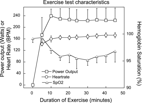
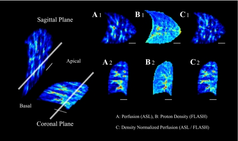
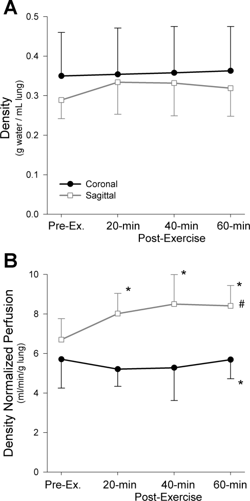
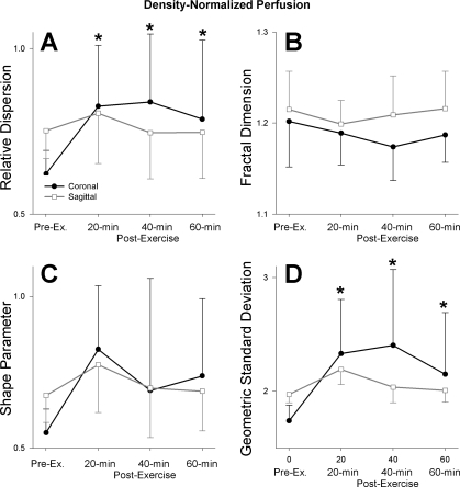
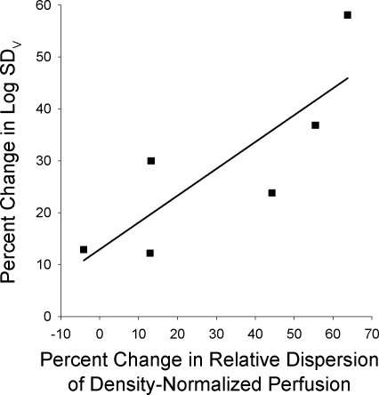
Exercise presents a considerable stress to the pulmonary system and ventilation-perfusion (Va/Q) heterogeneity increases with exercise, affecting the efficiency of gas exchange. In particular, prolonged heavy exercise and maximal exercise are known to increase Va/Q heterogeneity and these changes persist into recovery. We hypothesized that the spatial heterogeneity of pulmonary perfusion would be similarly elevated after prolonged exercise. To test this, athletic subjects (n = 6, Vo(2max) = 61 ml. kg(-1).min(-1)) with exercising Va/Q heterogeneity previously characterized by the multiple inert gas elimination technique (MIGET), performed 45 min of cycle exercise at approximately 70% Vo(2max). MRI arterial spin labeling measures of pulmonary perfusion were acquired pre- and postexercise (at 20, 40, 60 min post) to quantify the spatial distribution in isogravitational (coronal) and gravitationally dependent (sagittal) planes. Regional proton density measurements allowed perfusion to be normalized for density and quantified in milliliters per minute per gram. Mean lung density did not change significantly in either plane after exercise (P = 0.19). Density-normalized perfusion increased in the sagittal plane postexercise (P =or <0.01) but heterogeneity did not (all P >or= 0.18), likely because of perfusion redistribution and vascular recruitment. Density-normalized perfusion was unchanged in the coronal plane postexercise (P = 0.66), however, perfusion heterogeneity was significantly increased as measured by the relative dispersion [RD, pre 0.62(0.07), post 0.82(0.21), P < 0.0001] and geometric standard deviation [GSD, pre 1.74(0.14), post 2.30(0.56), P < 0.005]. These changes in heterogeneity were related to the exercise-induced changes of the log standard deviation of the ventilation distribution, an MIGET index of Va/Q heterogeneity (RD R(2) = 0.68, P < 0.05, GSD, R(2) = 0.55, P = 0.09). These data are consistent with but not proof of interstitial pulmonary edema as the mechanism underlying exercise-induced increases in both spatial perfusion heterogeneity and Va/Q heterogeneity.


Figures

Fig. 1.
Fig. 1.
Characteristics of the exercise test. Data are mean(SD). SpO2, peripheral arterial hemoglobin oxygen saturation as measured by pulse oximetry. BPM, beats/min.
Fig. 2.
Fig. 2.
This figure was previously published in Ref. . Magnetic resonance (MR) images of a representative subject in the sagittal and coronal planes of the lung. Images to the left depict the relationship of the imaging planes to one another, with the thick, long, white bar denoting the intersection of the 2 planes in the right lung. The smaller, thin, white bar illustrates a 3-cm scale on the images. Images A1, B1, and C1 are taken in the sagittal plane; the images A2, B2, C2 are from the coronal plane. Distributions of pulmonary perfusion measured by arterial spin-labeling (ASL) are shown in A1 and A2; signal intensity is proportional to perfusion (ml blood·min−1·ml lung−1). B1 and B2 depict a map of lung density using fast low-angle shot (FLASH) measures of proton density, where lung density is proportional to signal intensity in units of g water/cm3 lung. The division of the ASL perfusion images by the FLASH density images are demonstrated in C1 and C2. The signal intensity of these images reflects density-normalized pulmonary perfusion (ASL/FLASH; ml·min−1·g lung−1), which is perfusion per gram of lung water, including both blood and tissue.
Fig. 3.
Fig. 3.
Mean density (A) and density-normalized perfusion (B) in both the coronal and sagittal imaging planes. No significant differences in mean density were observed pre- or postexercise (P = 0.19) or between imaging planes (P = 0.32). The density-normalized perfusion in the saggital plane was significantly higher than the coronal plane (#P < 0.001). Additionally, there was a significant interaction noted between imaging plane and time postexercise (P < 0.05) accounted for by significant increases in the density-normalized perfusion postexercise at all time points (*P < 0.01) in the saggital plane, whereas the coronal density-normalized perfusion did not change significantly after exercise (P = 0.66).
Fig. 4.
Fig. 4.
Effect of exercise on heterogeneity of density-normalized pulmonary perfusion as measured by relative dispersion (A), fractal dimension (B), shape parameter (C), and geometric standard deviation (D). There was a significant interaction between the imaging plane and time postexercise in the relative dispersion (A; P < 0.0001) reflecting significant increases in the coronal plane postexercise (*P < 0.005) but not in the sagittal plane (P = 0.56). The fractal dimension (B) was not significantly different between imaging planes (P = 0.14), nor was there a significant difference after exercise (P = 0.09). The shape parameter (C) was not significantly different between imaging planes (P = 0.91) or after exercise (P = 0.08). The geometric standard deviation (D) showed a significant interaction between image plane and time postexercise (P < 0.0005), which was accounted for by a significant increase in the geometric standard deviation in the coronal plane postexercise (*P < 0.005), while the changes in the sagittal plane were not significant (P = 0.18).
Fig. 5.
Fig. 5.
Linear relationship between percent change from pre- to postexercise in the relative dispersion of density-normalized perfusion in the coronal plane vs. percent change in ventilation-perfusion heterogeneity from rest to 90% of maximal exercise. Ventilation perfusion heterogeneity was measured by the logarithm of the standard deviation of the ventilation distribution, Log SDV, from previously acquired multiple inert gas elimination technique data. There was a significant positive relationship (P < 0.05, R2 = 0.675).

References

    1. Anholm JD, Milne EN, Stark P, Bourne JC, Friedman P. Radiographic evidence of interstitial pulmonary edema after exercise at altitude. J Appl Physiol 86: 503–509, 1999. - PubMed
    1. Arai TJ, Henderson AC, Dubowitz DJ, Levin DL, Friedman PJ, Buxton RB, Prisk GK, Hopkins SR. Hypoxic pulmonary vasoconstriction does not contribute to pulmonary blood flow heterogeneity in normoxia in normal supine humans. J Appl Physiol 106: 1057–1064, 2009. - PMC - PubMed
    1. Beaver WL, Wasserman K, Whipp BJ. A new method for detecting anaerobic threshold by gas exchange. J Appl Physiol 60: 2020–2027, 1986. - PubMed
    1. Bernard SL, Glenny RW, Erickson HH, Fedde MR, Polissar N, Basaraba RJ, Hlastala MP. Minimal redistribution of pulmonary blood flow with exercise in racehorses. J Appl Physiol 81: 1062–1070, 1996. - PubMed
    1. Bolar DS, Levin DL, Hopkins SR, Frank LF, Liu TT, Wong EC, Buxton RB. Quantification of regional pulmonary blood flow using ASL-FAIRER. Magn Reson Med 55: 1308, 2006. - PubMed

Publication types

LinkOut - more resources