. 2009 Nov 6;284(45):31249-59.
doi: 10.1074/jbc.M109.041830. Epub 2009 Sep 10.

Molecular identification and functional characterization of Arabidopsis thaliana mitochondrial and chloroplastic NAD+ carrier proteins

Affiliations

Molecular identification and functional characterization of Arabidopsis thaliana mitochondrial and chloroplastic NAD+ carrier proteins

Ferdinando Palmieri et al. J Biol Chem. .

Abstract

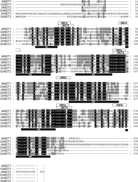
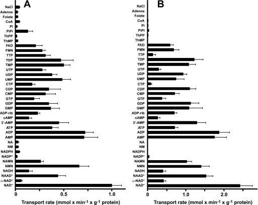
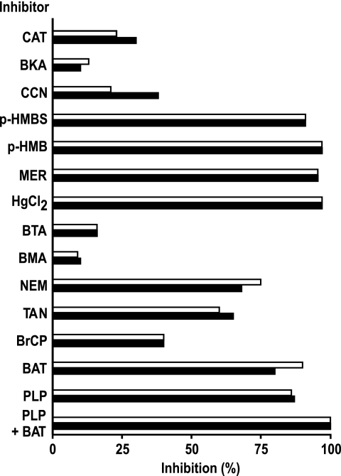
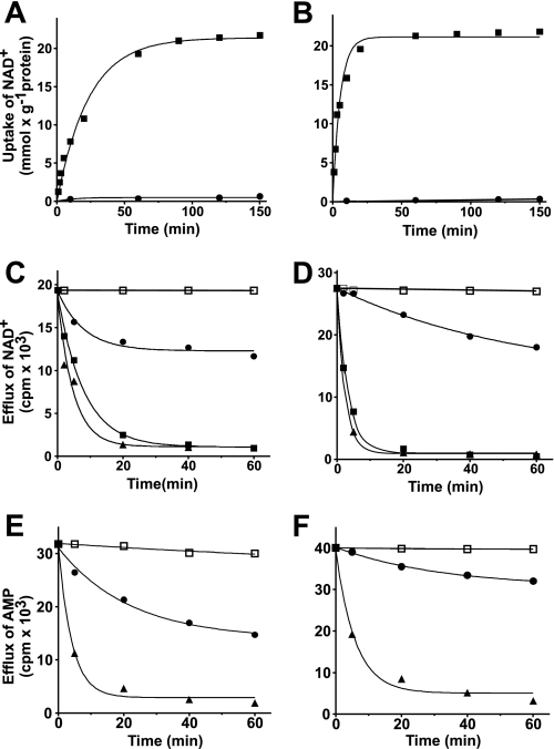
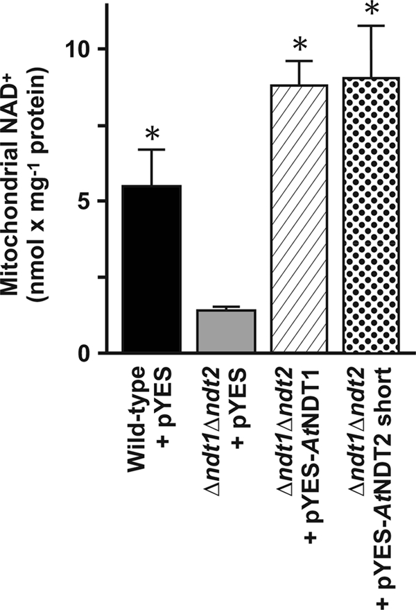
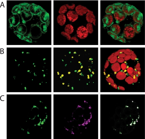
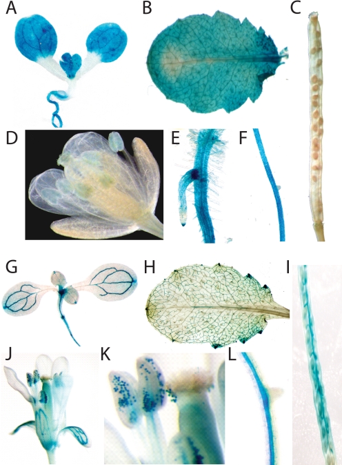
The Arabidopsis thaliana L. genome contains 58 membrane proteins belonging to the mitochondrial carrier family. Two mitochondrial carrier family members, here named AtNDT1 and AtNDT2, exhibit high structural similarities to the mitochondrial nicotinamide adenine dinucleotide (NAD(+)) carrier ScNDT1 from bakers' yeast. Expression of AtNDT1 or AtNDT2 restores mitochondrial NAD(+) transport activity in a yeast mutant lacking ScNDT. Localization studies with green fluorescent protein fusion proteins provided evidence that AtNDT1 resides in chloroplasts, whereas only AtNDT2 locates to mitochondria. Heterologous expression in Escherichia coli followed by purification, reconstitution in proteoliposomes, and uptake experiments revealed that both carriers exhibit a submillimolar affinity for NAD(+) and transport this compound in a counter-exchange mode. Among various substrates ADP and AMP are the most efficient counter-exchange substrates for NAD(+). Atndt1- and Atndt2-promoter-GUS plants demonstrate that both genes are strongly expressed in developing tissues and in particular in highly metabolically active cells. The presence of both carriers is discussed with respect to the subcellular localization of de novo NAD(+) biosynthesis in plants and with respect to both the NAD(+)-dependent metabolic pathways and the redox balance of chloroplasts and mitochondria.


Figures

FIGURE 1.
FIGURE 1.
Alignment of the predicted amino acid sequences of AtNDT1 and AtNDT2 with NDT homologues. The residues identical or similar among all family members are indicated by black shading, and the residues conserved by three proteins are indicated by gray shading. Solid black bars underline six putative membrane-spanning helices (H1–H6). Three conserved mitochondrial energy transfer signatures (mitochondrial energy transfer signature = PX(DE)XX(RK)X(RK)) after each odd membrane-spanning helix are marked by white bars. Boxes above the sequences indicate part a and part b of the 3-fold repeated signature motive (synthetic minimal medium) characteristic of the MCF proteins. The numbers indicate the amino acid positions. AtNDT1, nicotinamide adenine dinucleotide transporter 1 from A. thaliana (NCB accession no. AAC62861); AtNDT2, nicotinamide adenine dinucleotide transporter2 from A. thaliana (NCB accession no. AAP42759); OsNDT1, nicotinamide adenine dinucleotide transporter1 from O. sativa (NCB accession no. AAV43947.1); OsNDT2, nicotinamide adenine dinucleotide transporter2 from O. sativa (NCB accession no. BAD73272.1); ScNDT1, nicotinamide adenine dinucleotide transporter1 from S. cerevisiae (NCB accession no. NP_012260); ScNDT2, nicotinamide adenine dinucleotide transporter2 from S. cerevisiae (NCB accession no. NP_010910).
FIGURE 2.
FIGURE 2.
Dependence on internal substrate of the transport properties of proteoliposomes reconstituted with recombinant AtNDT1 (A) and AtNDT2 (B). Proteoliposomes were preloaded internally with various substrates (each concentration 10 mm). Transport was started by adding 0.6 mm and 0.15 mm [3H]NAD+ for AtNDT1 and AtNDT2, respectively. The reaction time was 2 min (AtNDT1) and 45 s (AtNDT2). Data are the means ± S.D. of at least three independent experiments. Adenos, adenosine; ADP-rib, ADP-ribose; NA, nicotinic acid; ThMP, thiamine monophosphate; ThPP, thiamine pyrophosphate; NM, nicotinamide.
FIGURE 3.
FIGURE 3.
Effect of inhibitors on the [3H]NAD+/NAD+ exchange by reconstituted AtNDT1 and AtNDT2. Proteoliposomes were preloaded internally with 10 mm NAD+. Transport was initiated by adding 0.6 and 0.15 mm [3H]NAD+ for AtNDT1 (white bars) or AtNDT2 (black bars), respectively. The reaction time was 2 min (AtNDT1) and 45 s (AtNDT2), respectively. Thiol reagents were added 2 min before the labeled substrate; the other inhibitors were added together with the labeled substrate. The final concentrations of the inhibitors were 20 mm (PLP, pyridoxal 5′-phosphate; BAT, bathophenanthroline), 0.2 mm (MER, mersalyl; p-HMB, p-hydroxylmercuribenzoate; p-HMBS, p-hydroxymercuribenzensulfonate), 25 μm HgCl2, 2 mm (BM, butylmalonate; BTA, 1,2,3-benzenetricarboxylate), 0.3 mm (BrCP, bromcresol purple), 1 mm (NEM, N-ethylmaleimide; CCN, α-cyano-4-hydroxycinnamate), 0.2% (TAN, tannic acid), and 10 μm (BKA, bongkrekic acid; CAT, carboxyatractyloside). The extents of inhibition (%) for each carrier from a representative experiment are given.
FIGURE 4.
FIGURE 4.
Kinetics of exchange reactions catalyzed by AtNDT1 and AtNDT2. Proteoliposomes were reconstituted with the recombinant AtNDT1 (A, C, and E) or AtNDT2 (B, D, and F). A and B, 0.6 mm [3H]NAD was added to proteoliposomes containing 10 mm NAD+ (■) or 10 mm NaCl (●). Similar results were obtained in three independent experiments. C, D, E, and F, the internal substrate of proteoliposomes (2 mm NAD+ in C and D or 2 mm AMP in E and F) was labeled with [3H]NAD (C and D) or [14C]AMP (E and F) by carrier-mediated exchange equilibration. After removal of the external substrate by Sephadex G-75, the efflux of [3H]NAD (C and D) or [14C]AMP (E and F) was started by adding buffer A alone (●), 5 mm NAD+ (▴), 5 mm AMP (■) or 5 mm NAD+, 20 mm pyridoxal 5′-phosphate, and 20 mm bathophenanthroline in buffer A (□). Similar results were obtained in three independent experiments. S.E. was always below 8% of the given value.
FIGURE 5.
FIGURE 5.
Effect of AtNDT1 and AtNDT2 expression on the mitochondrial NAD+ content of the Δndt1Δndt2 double mutant yeast cells. Mitochondria were isolated from wild-type + pYES2 (black bar), Δndt1Δndt2 + pYES2 (gray bar), Δndt1Δndt2 + pYES-AtNDT1 (hatched bar), and Δndt1Δndt2 + pYES-AtNDt2 short (dotted bar) cells grown on synthetic complete medium supplemented with 2% ethanol and 0.4% galactose. Data are the means ± S.D. of at least three independent experiments. The asterisk indicates significant differences in the mitochondrial NAD+ content as compared with that of Δndt1Δndt2 + pYES cells.
FIGURE 6.
FIGURE 6.
Localization of AtNDT1- and AtNDT2-GFP proteins in tobacco leaf protoplasts. Tobacco protoplasts were prepared as given under “Experimental Procedures” and transformed using polyethylene glycol. Fluorescence was visualized after 18 h of incubation by fluorescence microscopy. Chloroplasts were visualized by their autofluorescence. A, shown is localization of AtNDT1-GFP. B, shown are localization of AtNDT2-GFP and cotransformation with SKL-DSred. C, shown is the localization of AtNDT2-GFP and coincubation with the Mito Tracker dye.
FIGURE 7.
FIGURE 7.
Histochemical localization of GUS expression under control of the Atndt1 and Atndt2 promoter in A. thaliana. Plants were grown as described under “Experimental Procedures.” A, shown is Atndt1-promoter-GUS activity in a 10-day-old seedling. B, shown is Atndt1-promoter-GUS activity in a juvenile leaf from a 7-week-old plant. C, shown is Atndt1-promoter-GUS activity in developing siliques. D, shown is Atndt1-promoter-GUS activity in flowers. E and F, shown are Atndt1-promoter-GUS activity in root tips and root branches. G, shown is Atndt2-promoter-GUS activity in 2-week-old seedlings. H, shown is Atndt2-promoter-GUS activity in a mature leaf. I, shown is Atndt2-promoter-GUS activity in developing siliques. J, shown is Atndt2-promoter-GUS activity in flowers. K, shown is Atndt2-promoter-GUS activity in anthers. L, shown is Atndt2-promoter-GUS activity in roots.

References

    1. Heldt H. W. (2005) Plant Biochemistry, Elsevier Academic Press, Burlington, MA
    1. Roux S. J., Steinebrunner I. (2007) Trends Plant Sci. 12, 522–527 - PubMed
    1. Lehninger A. L., Nelson D. L., Cox M. M. (1994) Prinzipien der Biochemie, Spektrum, Akad. Verlag, Heidelberg, Berlin, Oxford
    1. Klingenberg M. (2008) Biochim. Biophys. Acta 1778, 1978–2021 - PubMed
    1. Strotmann H., Berger S. (1969) Biochem. Biophys. Res. Com. 35, 20–26 - PubMed

Publication types

MeSH terms