. 2009 Nov 4;28(21):3353-65.
doi: 10.1038/emboj.2009.260. Epub 2009 Sep 10.

Mechanism for the definition of elongation and termination by the class II CCA-adding enzyme

Affiliations

Mechanism for the definition of elongation and termination by the class II CCA-adding enzyme

Yukimatsu Toh et al. EMBO J. .

Abstract

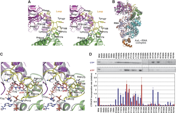
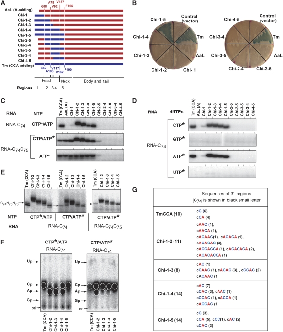
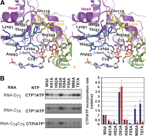
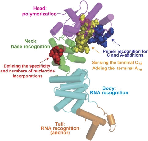
The CCA-adding enzyme synthesizes the CCA sequence at the 3' end of tRNA without a nucleic acid template. The crystal structures of class II Thermotoga maritima CCA-adding enzyme and its complexes with CTP or ATP were determined. The structure-based replacement of both the catalytic heads and nucleobase-interacting neck domains of the phylogenetically closely related Aquifex aeolicus A-adding enzyme by the corresponding domains of the T. maritima CCA-adding enzyme allowed the A-adding enzyme to add CCA in vivo and in vitro. However, the replacement of only the catalytic head domain did not allow the A-adding enzyme to add CCA, and the enzyme exhibited (A, C)-adding activity. We identified the region in the neck domain that prevents (A, C)-adding activity and defines the number of nucleotide incorporations and the specificity for correct CCA addition. We also identified the region in the head domain that defines the terminal A addition after CC addition. The results collectively suggest that, in the class II CCA-adding enzyme, the head and neck domains collaboratively and dynamically define the number of nucleotide additions and the specificity of nucleotide selection.


Conflict of interest statement

The authors declare that they have no conflict of interest.

Figures

Figure 1
Figure 1
Crystal structures of T. maritima CCA-adding enzyme (TmCCA) and its complexes with CTP and ATP. (A) Overall structure of form I of apo TmCCA. The head, neck, body and tail domains of TmCCA are coloured magenta, green, cyan and orange, respectively. The loop between β6 and α5 in the head domain (amino acids 102–121) is disordered and is highlighted by a circle. (B) Superposition of the TmCCA forms I and II structures. The main chains of TmCCA forms I and II are coloured green and magenta, respectively. The loop region visible in the form II structure is circled. (C) CTP (upper panel) and ATP (lower panel) recognition by the TmCCA active site of the form I crystals. CTP and ATP are coloured blue and red, respectively. (D) Superposition of the active sites of complex structures with CTP and ATP. The CTP and ATP complex structures are coloured cyan and magenta, respectively. CTP and ATP are coloured blue and red, respectively, as in (C).
Figure 2
Figure 2
Structure of the flexible loop in the catalytic head domain of T. maritima CCA-adding enzyme (TmCCA). (A) Structure of the catalytic head domain of crystal form II of TmCCA. The loop region (amino-acid residues 102–121) is coloured yellow. (B) The structure of crystal form II TmCCA (coloured as in Figure 1A) was superposed on the structure of Aquifex aeolicus A-adding enzyme complexed with tRNA lacking A76 and an incoming ATP analog (AaL–RNA complex, coloured gray, PDB code: 1VFG; Tomita et al, 2004). The head and neck domains of the two enzymes are superposed. (C) The detailed stereo view of the catalytic pocket of the superposition in (B). The loop region in the head domain is coloured yellow. ATP is coloured red. (D) In vitro relative CMP and AMP incorporation rates by mutant TmCCA variants. Asterisks indicate the α-32P-nucleotide used in the assays. The blue and red bars in the graph indicate the relative CMP and AMP incorporation rates, respectively. The CMP and AMP incorporation rates into mini-C74 and mini-C74C75 by the wild-type TmCCA are defined as 1.0. The bars in the graph are the standard deviations of more than three independent experiments.
Figure 3
Figure 3
Compatibility of the loop regions between T. maritima CCA-adding enzyme and A. aeolicus A-adding enzyme. (A) Representation of the chimaeric proteins of T. maritima CCA-adding enzyme (Tm; blue), A. aeolicus A-adding enzyme (AaL; red) and A. aeolicus CC-adding enzyme (AaS, green) (upper). The amino-acid sequences of the loop regions of TmCCA (amino-acid residues 101–121, coloured blue), Aquifex aeolicus A-adding enzyme (amino-acid residues 76–96, coloured red) and CC-adding enzyme (amino-acid residues 114–124, coloured green) (lower). The catalytic residues are underlined. (B) In vivo conditional suppression assays by the chimaeric enzymes. E. coli strain CA224cca, carrying the plasmid encoding an amber suppressor tRNA gene [pSup(CGGOH)], was transformed by the pMW118 plasmid either with no insert (control), the Tm, Tm-AaL-loop and Tm-AaS-loop genes (upper left plate), the AaL, AaL-Tm-loop and AaL-AaS-loop genes (upper right plate), or the AaS, AaS-Tm-loop and AaS-AaL-loop genes (lower plate). The transformants were inoculated on LB plates containing ampicillin, chloramphenicol, tetracycline, IPTG and X-gal. (C) In vitro CCA-adding assays by the recombinant chimaeric proteins in (A). CMP incorporation into mini-C74 in the presence of 32P-CTP and unlabelled ATP (upper panel), and AMP incorporation into mini-C74C75 in the presence of 32P-ATP (lower panel). Asterisks indicate the α-32P-nucleotide used in the assays.
Figure 4
Figure 4
Generation of the CCA-adding enzyme from the A-adding enzyme in vivo and in vitro. (A) Representations of the chimaeric TmCCA–AaL enzymes. The protein regions of Aquifex aeolicus A-adding enzyme (AaL) and T. maritima CCA-adding enzyme (TmCCA) are coloured red and blue, respectively. The amino-acid sequence numbering of AaL and TmCCA is coloured red and blue, respectively. (B) In vivo amber suppression assays by chimaeric TmCCA–AaL enzymes in (A). The transformants were inoculated on LB plates as in Figure 3. (C) In vitro CCA-adding assays using the recombinant chimaeric TmCCA–AaL variants in (A). CMP incorporation into mini-C74 in the presence of 32P-CTP and unlabelled ATP (upper panel). AMP incorporation into mini-C74C75 in the presence of 32P-ATP and unlabelled CTP (middle panel) and in the presence of 32P-ATP (lower panel). (D) CMP or AMP incorporation by chimaeric enzymes into mini-C74 RNA in the presence of all four nucleotides. Neither GMP nor UMP is incorporated by the chimaeric enzymes. (E) Separation of reaction products at one nucleotide resolution. CMP or AMP incorporation into RNA ending with C74 (left) and into RNA ending with C74C75 (right) by TmCCA and chimaeric enzymes (Chi-1-2, Chi-1-3, Chi-1-4 and Chi-1-5). The arrows in the gel margins indicate the product with the expected CCA end. (F) Neighbouring nucleotide analysis of 32P-labelled products in (E) by thin-layer chromatography. Tm(CCA), Chi-1-2, Chi-1-3, Chi-1-4 and Chi-1-5 indicate RNase T2 hydrolysates of 32P-labelled products in the gels in the left part of (E) (left, CTP-labelled product) and middle part of (E) (right, ATP-labelled product). (G) Nucleotide sequences of 3′-terminal regions of reaction products generated by chimaeric enzymes. The total number of the clones analysed (left columns) and the number of the clones with the respective sequences (right columns) are shown in parentheses. Asterisks in (C), (D), (E) and (F) indicate the α-32P-nucleotide used in the assays.
Figure 5
Figure 5
Hydrogen bonds in the neck domain involved in the correct CCA addition. (A) Detailed representation of chimaeras Chi-1-4 and Chi-1-5 in Figure 4A. The protein regions of Aquifex aeolicus A-adding enzyme (AaL) and T. maritima CCA-adding enzyme (TmCCA) are coloured red and blue, respectively. The amino-acid sequences of region-5 of AaL and TmCCA are shown. The different amino-acid residues are underlined. (B) In vivo amber suppression assays by Chi-1-4 bearing the Glu185Gln186 sequence in region 5 (left plate). The transformants were inoculated on LB plates, as in Figure 3. In vitro CCA-adding assays by the recombinant protein Chi-1-4 EQ (right). The CMP incorporation into mini-C74 (upper panel) and the AMP incorporation into mini-C74C75 (lower panel). Asterisks indicate the α-32P-nucleotide used in the assays. Arrows at the gel margin indicate the RNA products with the CCA end. (C) Structures of the backside of the neck domains of TmCCA (left) and AaL (middle). The hydrogen bonds between α9 and α13 of TmCCA are depicted by dashed lines. A model structure of AaL with the mutations of Ala160Gly161 to Glu160Gln161 (AaL-EQ model: right).
Figure 6
Figure 6
Involvement of the β-turn in the head domain in the CCA addition by T. maritima CCA-adding enzyme. (A) Stereo views of the superposition of the form II TmCCA structure on the structure of A. aeolicus A-adding enzyme complexes with the RNA primer and ATP, as in Figure 2C. The protein structure of A. aeolicus A-adding enzyme is not shown. Only the catalytic domain of TmCCA is shown. The β-turn (amino-acid residues 81–87) of TmCCA is coloured blue. (B) In vitro relative CMP and AMP incorporation rates by mutant TmCCA variants. The CMP and AMP incorporation into mini-D73, mini-C74 and mini-C74C75 by the wild-type TmCCA (left gel panels). The blue and red bars in the graph indicate the relative CMP and AMP incorporation rates into mini-C74 and mini-C74C75, respectively. The bars in the graph are the standard deviations of more than three independent experiments. As the incorporation of CMP into mini-D73 represents the CMP incorporation at positions 74 and 75, the quantification of the CMP incorporation rate was performed only for the CMP incorporation into mini-C74.
Figure 7
Figure 7
Functional elements for the CCA-adding reaction. The overall structure of form II of TmCCA. The head, neck, body and tail domains of TmCCA are coloured magenta, green, cyan and orange, respectively. The loop in the head domain (presented as yellow spheres) is involved in the recognition of the terminal C75 of the primer and in A76 incorporation. The hydrogen bond between the two helices in the neck domain defines the specificity and the number of nucleotide incorporations into the RNA. Amino-acid residues forming hydrogen bonds are depicted as red spheres. The β-turn in the catalytic domain (represented as blue spheres) is involved in the recognition of 3′ end of the primer RNA for CMP and AMP incorporations.

Similar articles

Cited by

References

    1. Adams PD, Grosse-Kunstleve RW, Hung LW, Ioerger TR, McCoy AJ, Moriarty NW, Read RJ, Sacchettini JC, Sauter NK, Terwilliger TC (2002) PHENIX: building new soft warefor automated crystallographic structure determination. Acta Crystallogr D Biol Crystallogr 58: 1948–1954 - PubMed
    1. Augustin MA, Reichert AS, Betat H, Huber R, Mörl M, Steegborn C (2003) Crystal structure of the human CCA-adding enzyme: insights into template-independent polymerization. J Mol Biol 328: 985–994 - PubMed
    1. Bralley P, Chang SA, Jones GH (2005) A phylogeny of bacterial RNA nucleotidyltransferases: Bacillus halodurans contains two tRNA nucleotidyltransferases. J Bacteriol 187: 5927–5936 - PMC - PubMed
    1. Brunger AT, Adams PD, Clore GM, DeLano WL, Gros P, Grosse-Kunstleve RW, Jiang JS, Kuszewski J, Nilges M, Pannu NS, Read RJ, Rice LM, Simonson T, Warren GL (1998) Crystallography and NMR system: a new software suite for macromolecular structure determination. Acta Crystallogr D 54: 905–921 - PubMed
    1. Cho HD, Verlinde CL, Weiner AM (2005) Archaeal CCA-adding enzymes: central role of a highly conserved beta-turn motif in RNA polymerization without translocation. J Biol Chem 280: 9555–9566 - PubMed

Publication types

MeSH terms