. 2009 Oct 21;28(20):3171-84.
doi: 10.1038/emboj.2009.256. Epub 2009 Sep 10.

A new plant protein interacts with eIF3 and 60S to enhance virus-activated translation re-initiation

Affiliations

A new plant protein interacts with eIF3 and 60S to enhance virus-activated translation re-initiation

Odon Thiébeauld et al. EMBO J. .

Abstract

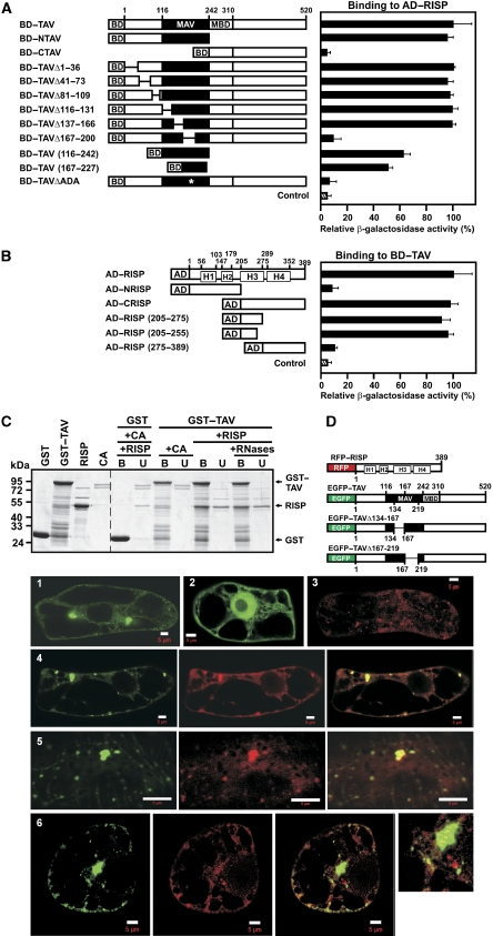
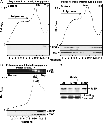
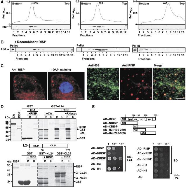
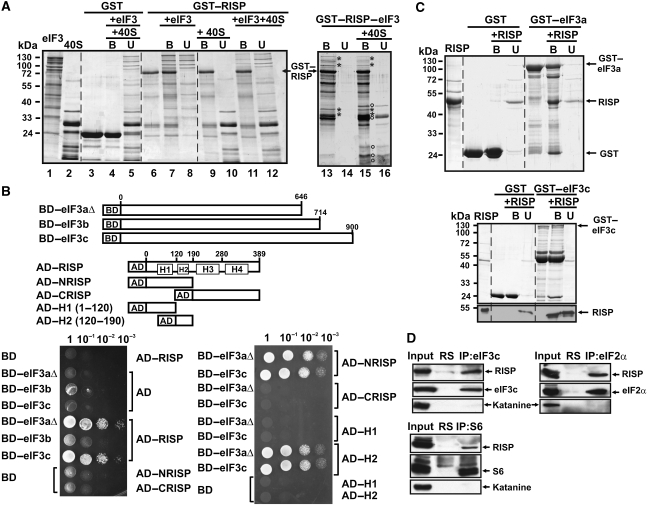
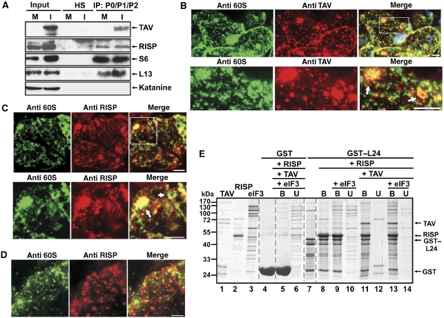
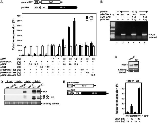
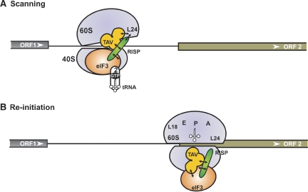
The plant viral re-initiation factor transactivator viroplasmin (TAV) activates translation of polycistronic mRNA by a re-initiation mechanism involving translation initiation factor 3 (eIF3) and the 60S ribosomal subunit (60S). QJ;Here, we report a new plant factor-re-initiation supporting protein (RISP)-that enhances TAV function in re-initiation. RISP interacts physically with TAV in vitro and in vivo. Mutants defective in interaction are less active, or inactive, in transactivation and viral amplification. RISP alone can serve as a scaffold protein, which is able to interact with eIF3 subunits a/c and 60S, apparently through the C-terminus of ribosomal protein L24. RISP pre-bound to eIF3 binds 40S, suggesting that RISP enters the translational machinery at the 43S formation step. RISP, TAV and 60S co-localize in epidermal cells of infected plants, and eIF3-TAV-RISP-L24 complex formation can be shown in vitro. These results suggest that RISP and TAV bridge interactions between eIF3-bound 40S and L24 of 60S after translation termination to ensure 60S recruitment during repetitive initiation events on polycistronic mRNA; RISP can thus be considered as a new component of the cell translation machinery.


Conflict of interest statement

The authors declare that they have no conflict of interest.

Figures

Figure 1
Figure 1
Association of re-initiation supporting protein (RISP) with transactivator viroplasmin (TAV) and mapping of their interaction domains. (A) Interaction between TAV and its deletion mutants fused to the Gal4 binding domain (BD) and RISP fused to Gal4 activation domain (AD) in the yeast two-hybrid system was quantified by measuring β-galactosidase activity. The highest value of β-galactosidase activity in diploids transformed with both full-length constructs was taken as 100% (12 Miller units). MAV, minimal segment of TAV; and MBD, multiple protein-binding domain. (B) Quantification of interactions between RISP and its deletion mutants fused to Gal4 AD and BD-TAV. H1–H4 predicted coiled-coil domains. (C) GST and GST–TAV bound to glutathione beads were incubated with either purified recombinant RISP or Conalbumin (CA, 75 kDa). Lanes (+ RNases) show the experiment carried out in the presence of an RNase cocktail. The beads were washed, and the unbound (U) and bound (B) fractions were analyzed by SDS–PAGE followed by Coomassie blue staining. Right panel, Interactions of RISP with GST or GST–TAV; and left panel, purified GST, GST–TAV and RISP. (D) Schematic representation of full-length RISP fused to the C-terminus of RFP, and full-length TAV (or truncated versions) fused to the C-terminus of enhanced green fluorescent protein (EGFP). Panels 1–6: Imaging fluorescence assays showing tobacco BY-2 cells transiently expressing EGFP–TAV (green, 1), EGFP alone (green, 2), or RFP-RISP (red, 3). 4 Left: EGFP–TAV, central: red fluorescent protein (RFP)–RISP, and right: merged. 5 Left: EGFP–TAVΔ134–167, central: RFP–RISP, right: merged. 6 Left: EGFP–TAVΔ167–219, central: RFP–RISP, right: merged; far right panel: high magnification image of part of the cell. Scale bars, 5 μm.
Figure 2
Figure 2
Re-initiation supporting protein (RISP) and transactivator viroplasmin (TAV) accumulate in polyribosomes during viral infection. (A, B) Ribosomal profiles of polyribosomes and ribosomal species from healthy (left) and Cauliflower mosaic virus (CaMV)-infected (right) turnip plants (A) untreated and (B) treated with 30 mM EDTA. A, B show the UV profile of the gradient with 40S and 60S, monosomes (80S) and polysomes indicated are shown. 1 ml aliquot fractions were either precipitated with 10% TCA and analyzed by SDS–PAGE and immunoblotting using polyclonal antibodies against TAV and RISP (lower panels); or analyzed by agarose gel electrophoresis (upper panel in B). Positions of 18S and 28S rRNAs are indicated. (C) Immunoblotting using polyclonal antibodies against RISP of extracts isolated from Arabidopsis (At), healthy (Turnip −) and CaMV-infected (Turnip +), turnip plants and recombinant RISP expressed in Escherichia coli. Loading controls are shown (lower panel).
Figure 3
Figure 3
Re-initiation supporting protein (RISP) binds the 60S ribosomal protein L24 and co-sediments with 60S and 80S. (A) RISP co-sediments with 80S (left), and 60S (middle) but not with 40S (right) ribosomes. Lower panels: immunostaining of gradient fractions with antibodies against RISP. (B) 80S, 60S and 40S were incubated with recombinant RISP at approximately 1:1 molar ratio before being subjected to sucrose density gradient centrifugation as in (A) followed by western blot analysis with antibodies against RISP. (C) Left panels: Immunofluorescence assays showing localization of RISP (red) within cytoplasm of BY-2 cells. Nucleus was stained with DAPI (blue); right panels: colocalization of endogenous 60S (green) and RISP (red) in BY-2 tobacco cells; only part of the cytoplasmic compartment is shown. Anti-60S and anti-RISP images were merged in the right-most panel. Scale bar, 5 μm. (D) RISP interacts with the C-terminal region of L24 in GST pull-down assay. RISP or the control protein CA was incubated with recombinant L24 fused to GST (GST–L24, upper panel). Lanes labelled +RNases show the experiment carried out in the presence of an RNase cocktail. RISP was mixed with either the N- or C-terminus of L24 fused to GST (GST–NL24 and GST–CL24; bottom panel) bound to glutathione beads. The beads were washed, and purified bound (B) and unbound (U) proteins were resolved by SDS–PAGE and stained with Coomassie blue. (E) Yeast two-hybrid interactions between BD–L24 and RISP and its deletion mutants fused to AD. Equal OD600 units and 1:1, 1:10 and 1:100 dilutions were spotted from left to right and incubated for 2 days.
Figure 4
Figure 4
Re-initiation supporting protein (RISP) binding to eIF3, through direct interaction with eIF3 subunits a and c, mediates its interaction with 40S. (A) GST–RISP binds to wheat eIF3 and 40S. Interactions of GST (lane 3), and GST–RISP (lane 6) bound to glutathione Sepharose 4B beads with eIF3 (lanes 7 and 8), 40S (lanes 9–10) or eIF3 and 40S (lanes 4–5 for GST and 11–12 for GST–RISP) are shown. The left panel (lanes 1 and 2) shows eIF3 and 40S. Interactions between the GST–RISP–eIF3 complex (lane 13) and 40S are shown in lanes 15–16 (right panel). Asterisks: characteristic eIF3 subunits; open circles: characteristic 40S proteins specifically co-precipitated with GST–RISP–eIF3. (B) The H2 domain of RISP interacts with eIF3aΔ and eIF3c in vivo. Schematic representation of BD–eIF3aΔ, BD–eIF3c and RISP, and its truncated versions fused to AD. Yeast two-hybrid analysis was carried out with RISP, AD–NRISP, AD–CRISP, and AD–H1 and AD–H2 against eIF3aΔ, eIF3b or eIF3c. Four dilutions of the transformation mixture are shown. (C) The H2 domain of RISP interacts with eIF3aΔ and eIF3c in GST pull-down assays. RISP was incubated with recombinant eIF3aΔ (upper panel) and eIF3c (bottom panel) fused to GST (GST–eIF3aΔ and GST–eIF3c) bound to glutathione beads. Purified proteins, and bound (B) and unbound (U) material were resolved by SDS–PAGE and stained with Coomassie blue. RISP distribution between the B and U fractions for GST–eIF3c was analyzed by western blot with polyclonal anti-RISP antibodies (lower panel). (D) Co-immunoprecipitation of RISP with eIF3c, or S6, or eIF2α. Arabidopsis suspension cultures were used for co-immunoprecipitation with either anti-eIF3c (upper left panel, or anti-eIF2α (upper right panel) or anti-S6 (bottom panel) antibodies. Each panel shows immunoblotting of RISP, eIF3c, S6, eIF2α and the control protein katanine present in input, normal rabbit serum (RS) and the entire immunoprecipitate (IP).
Figure 5
Figure 5
The re-initiation supporting protein (RISP)–transactivator viroplasmin (TAV) complex mediates contacts between 60S ribosomal protein L24 and eIF3. (A) Mock-inoculated (M) and cauliflower mosaic virus (CaMV)-infected (I) turnip plants were used for co-immunoprecipitation with anti-P0–P1–P2 antibodies. The panels show immunoblotting of RISP, TAV, S6 or L13 present in input, normal human serum (HS) and the entire immunoprecipitate (IP) using appropriate rabbit polyclonal antibodies. (B–D) Immunofluorescence assay showing colocalization of endogenous 60S and TAV (B) 60S and RISP in systemically CaMV-infected epidermal cells of leaves of B. rapa plants at 15 days post-inoculation (dpi; C) and 60S and RISP in mock-infected epidermal cells (D). Double-immunolabelling was carried out using anti-TAV and anti-P0–P1–P2 antibodies (anti-60S) (B) or anti-60S and anti-RISP antibodies (C, D). The nucleus was stained with DAPI (blue). The lower panels in (B) and (C) represent higher magnification images of the insets in the upper panels. In the merge, DAPI fluorescence is blue, TAV is red, 60S is green and RISP is red. Round-shaped structures are indicated by arrows. Scale bars, 5 μm. (E) GST pull-down assays. GST–L24 attached to glutathione Sepharose 4B beads (lane 7) was mixed either with RISP (lane 8), or RISP and eIF3 (lanes 9 and 10), or RISP and TAV (lanes 11–12), or RISP, TAV and eIF3 (lanes 13–14); purified TAV, RISP and eIF3 are shown in lanes 1, 2 and 3, respectively. Unbound (U) and bound (B) fractions were analyzed by SDS–PAGE followed by Coomassie blue staining.
Figure 6
Figure 6
Re-initiation supporting protein (RISP) participates in transactivator viroplasmin (TAV)-mediated transactivation in plant protoplasts. (A) Schematic diagram of the monocistronic chloramphenicol acetyltransferase (pmonoCAT) and dicistronic β-glucuronidase (pbiGUS) reporter constructs. Nicotiana plumbaginifolia protoplasts were co-transfected with the two reporter plasmids shown, as well as effector plasmids in the amounts indicated below the graph. All reporter and effector constructs were expressed under the control of the CaMV 35S promoter (35S). The amount of CAT (open bars) and GUS enzymatic activity (closed bars) synthesized in N. plumbaginifolia protoplasts is indicated. Results shown represent the means obtained in three independent experiments. Results are expressed as a percentage, with the amount of CAT and the enzymatic activity of GUS synthesized in protoplasts transfected with pTAV only being set as 100%. (B) Ability of TAV and TAVΔADA mutant to support Cauliflower mosaic virus (CaMV) replication. Semiquantitative reporter-targeted PCR analysis (25 cycles) of total LMW DNA from transfected protoplasts. Lane 1, mock-transfected protoplasts; lane 2, transfection with pE4Pin (10 μg) and pAATAV (wild-type; 4 μg); lane 3, pE4Pin (10 μg) alone; lane 4, pE4Pin (10 μg), pAA TAV (wild-type; 4 μg), pGW GAG (4 μg), pGW POL (2 μg); lane 5, pE4Pin (10 μg), pAA TAVΔADA (mutant, 4 μg); lane 6, pE4Pin (10 μg), pAA TAVΔADA (mutant, 4 μg); pGW GAG (4 μg), pGW POL (2 μg). LMW DNA, low molecular weight DNA, GAG, capsid protein precursor; POL, polyprotein with protease, reverse transcriptase, and RNase H activity. (C) Accumulation of RISP in wild-type and 134C07 Arabidopsis plants. Extracts from 0.1 g leaves were analyzed with rabbit polyclonal anti-RISP antibodies. (D) Kinetics of TAV and CP protein accumulation in CaMV infected wild-type and 134C07 plants. (E) Efficiency of TAV-mediated transactivation in mesophyll protoplasts prepared from wild-type (WT) and mutant (134C07) plants. GUS activity is shown as black bars; green fluorescent protein (GFP) accumulation was analyzed by western blot using anti-GFP antibodies. The data shown are the means of three independent assays, and error bars indicate s.d.
Figure 7
Figure 7
Proposed model of re-initiation supporting protein (RISP) function in 60S recruitment during virus-activated re-initiation. We propose the following scenario: during ORF1 elongation, the RISP–transactivator viroplasmin (TAV)–80S complex can be stabilized by transfer of TAV–RISP–eIF3 to the solvent surface of 60S through TAV binding to L18/L13. During termination, the TAV–RISP–eIF3 complex is relocated back to 40S to reconstruct a pre-initiation complex (PIC) competent for re-initiation. (A) RISP–TAV establishes a bridge between 40S-bound eIF3 and 60S through the ribosomal protein L24, preventing, for a short time, removal of 60S. During scanning, RISP bridges the relaxed 40S–60S interactions through contact with 40S-bound eIF3, while simultaneously stabilizing TAV–L24 contacts (open conformation of 80S). This open 80S conformation allows eIF3-bound 40S to continue scanning and search for a downstream start codon. (B) Codon–anticodon recognition and positioning of Met-tRNAiMet in the ribosomal P-site would then displace TAV and RISP from L24 followed by the formation of 80S ready for elongation. eIF3, TC, RISP, TAV, L24, 40S and 60S are indicated.

Similar articles

Cited by

References

    1. Alisch RS, Garcia-Perez JL, Muotri AR, Gage FH, Moran JV (2006) Unconventional translation of mammalian LINE-1 retrotransposons. Genes Dev 20: 210–224 - PMC - PubMed
    1. Ban N, Nissen P, Hansen J, Moore PB, Steitz TA (2000) The complete atomic structure of the large ribosomal subunit at 2.4 Å resolution. Science 289: 905–920 - PubMed
    1. Baronas-Lowell DM, Warner JR (1990) Ribosomal protein L30 is dispensable in the yeast Saccharomyces cerevisiae. Mol Cell Biol 10: 5235–5243 - PMC - PubMed
    1. Bonneville J-M, Sanfaçon H, Fütterer J, Hohn T (1989) Posttranscriptional transactivation in cauliflower mosaic virus. Cell 59: 1135–1143 - PubMed
    1. Bureau M, Leh V, Haas M, Geldreich A, Ryabova L, Yot P, Keller M (2004) P6 protein of Cauliflower mosaic virus, a translation reinitiator, interacts with ribosomal protein L13 from Arabidopsis thaliana. J Gen Virol 85: 3765–3775 - PubMed

Publication types

MeSH terms