. 2009 Oct 21;28(20):3256-68.
doi: 10.1038/emboj.2009.257. Epub 2009 Sep 10.

Pre-fibrillar alpha-synuclein variants with impaired beta-structure increase neurotoxicity in Parkinson's disease models

Affiliations

Pre-fibrillar alpha-synuclein variants with impaired beta-structure increase neurotoxicity in Parkinson's disease models

Damla Pinar Karpinar et al. EMBO J. .

Abstract

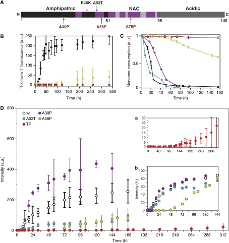
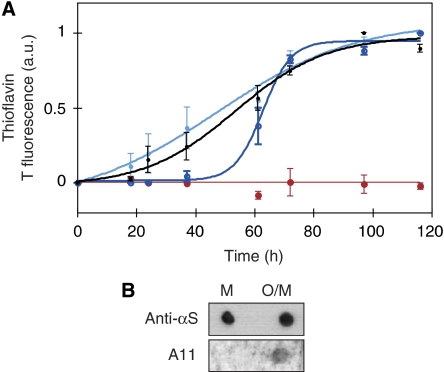
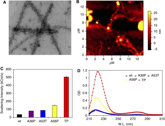
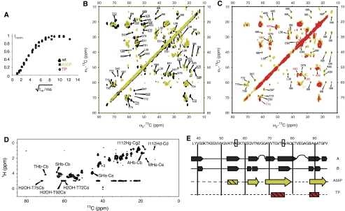
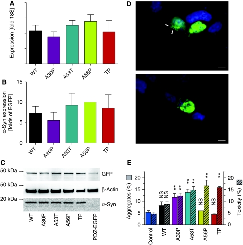
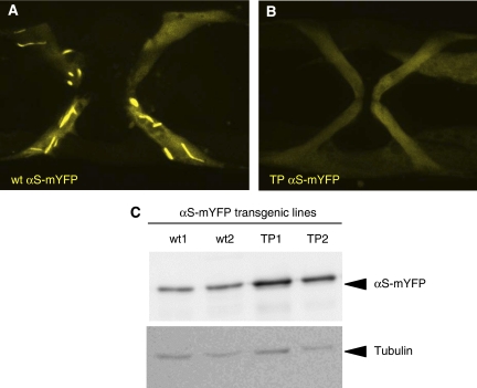
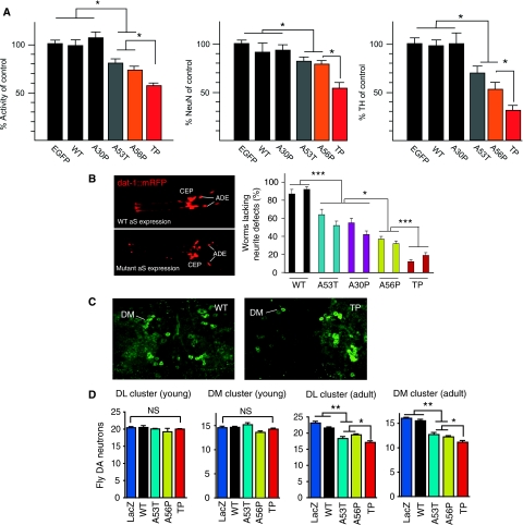
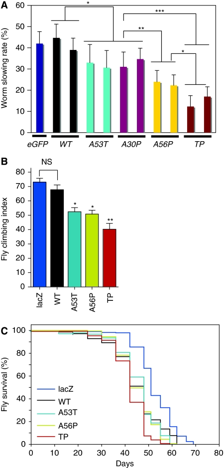
The relation of alpha-synuclein (alphaS) aggregation to Parkinson's disease (PD) has long been recognized, but the mechanism of toxicity, the pathogenic species and its molecular properties are yet to be identified. To obtain insight into the function different aggregated alphaS species have in neurotoxicity in vivo, we generated alphaS variants by a structure-based rational design. Biophysical analysis revealed that the alphaS mutants have a reduced fibrillization propensity, but form increased amounts of soluble oligomers. To assess their biological response in vivo, we studied the effects of the biophysically defined pre-fibrillar alphaS mutants after expression in tissue culture cells, in mammalian neurons and in PD model organisms, such as Caenorhabditis elegans and Drosophila melanogaster. The results show a striking correlation between alphaS aggregates with impaired beta-structure, neuronal toxicity and behavioural defects, and they establish a tight link between the biophysical properties of multimeric alphaS species and their in vivo function.


Conflict of interest statement

The authors declare that they have no conflict of interest.

Figures

Figure 1
Figure 1
Structure-based design mutants impair fibrillation of αS. (A) Functional domains of αS. Familial mutants (black) and structure-based design mutants (red) are labelled along the sequence. Regions involved in β-sheet formation in the fibril (Heise et al, 2005) are marked (purple). (B) Fibril formation of wt αS (black), A56P αS (yellow) and TP αS (red) followed by Thioflavin T fluorescence (ThT). Data for A30P, A76P, A30PA56P, A30PA76P are shown in Supplementary Figure S1. (C) Consumption of monomeric wt αS (black), A53T αS (cyan), A30P αS (purple), A56P αS (yellow) and TP αS (red) monitored through 1D 1H NMR spectroscopy. Drop in signal intensity is due to the formation of higher molecular weight aggregates not detectable by solution-state NMR. Errors were estimated from three independent aggregation assays. (D) Fibril formation of wt αS (black), A30P (purple), A53T (blue), A56P αS (yellow) and TP αS (red, also shown in Inset a at a different scale) followed by ThT fluorescence emission intensity. Inset b shows ThT intensity values of all but TP variants, each of them normalized by the maximal value observed along their aggregation reaction.
Figure 2
Figure 2
Aggregates formed by TP αS seed aggregation of wt αS. (A) Pre-fibrillar aggregates of TP αS seed fibril formation of wt αS. In an equimolar mixture of pre-aggregated TP αS with monomeric wt αS the lag time of aggregation is decreased (light blue) when compared with wt monomer alone (black). In contrast, in an equimolar mixture of monomeric TP αS and monomeric wt protein the lag time is increased (dark blue). Aggregation behaviour of monomeric TP αS alone is shown in red. Error bars represent mean±s.d. of three to four independent experiments. (B) Recognition of a mixture of oligomers and monomers of TP αS (O/M) but not monomeric TP αS (M) on nitrocellulose membrane by the A11 antibody. Anti-αS antibody shows comparable attachment to both monomer and oligomer.
Figure 3
Figure 3
Enhanced formation of soluble oligomers by αS variants. (A) Electron micrograph of TP αS solution in 50 mM HEPES, 100 mM NaCl (pH 7.4), 0.01% NaN3, incubated for 6 days at 37°C while being stirred at 200 r.p.m. The protein concentration was 0.8 mM, and the sample was diluted eightfold by buffer before EM imaging. (B) AFM image of TP αS solution. Conditions identical to (A). (C) Dynamic light scattering of αS variants, incubated for 11 days at the aggregation condition and then centrifuged briefly followed by the measurement of the supernatant. Data presented are average of three measurements, each consisting of 20 acquisitions of 20 s. (D) UV absorbance of the supernatant of aggregated αS variants after 11 days of incubation at the aggregation condition.
Figure 4
Figure 4
High-resolution solid-state NMR of late stage aggregates formed by αS variants. (A) Water accessibility of aggregates as probed by solid-state NMR. A 3-ms Gaussian pulse and a T2 filter containing two delays of 1 ms were used for selective water excitation (Ader et al, 2009). The cross polarization contact time was set to 700 μs. (B) Superposition of 2D 13C/13C correlation spectra of U-[13C,15N] A56P αS (yellow) and of wt αS (black). Correlations absent in the A56P mutant are underlined. Assignments correspond to values obtained for the A form of wt αS as reported in (Heise et al, 2005). (C) Superposition of 2D 13C/13C correlation spectra of U-[13C,15N] A56P αS (yellow) and of U-[13C,15N] TP αS (red). In (B) and (C), homonuclear mixing was achieved using a proton driven spin diffusion time of 20 ms (A56P) and 50 ms (TP), respectively. (D) 2D 1H/13C 1H-T2-filtered HETCOR spectrum of A56P αS. The spectrum was recorded at a magnetic field strength of 14 T, with a spinning speed of 8.33 kHz, at a sample temperature of 0°C. The T2 filter delay was 2 × 175 μs, the contact time was 3 ms. The spectrum was recorded without homonuclear decoupling during t1, 160 t1 increments and 128 scans per slice. (E) ssNMR-based secondary structure analysis for wt and mutant αS. Row 1 and 2 correspond to wt data reported previously (Heise et al, 2005). Hatched rectangles relate to protein regions in which β-strands are lost compared with wt αS or, in the case of TP, show strong dynamics/disorder. Arrows relate to β-strands that are preserved in A56P. Mutation sites are indicated by rectangular boxes, suggesting that A↔P mutation leads to partial (A56P) or almost complete (TP) suppression of β-strand formation in αS.
Figure 5
Figure 5
Aggregate formation and toxicity in HEK293T cells. (A) RT–PCR quantification of αS mRNA extracted 24 h after transfection. Bars represent αS mRNA relative to the ribosomal 18S subunit mRNA. (mean±s.e.m., n=3 independent experiments, no significant difference between different αS variants) (B) Quantification of the western blot bands (exemplified in C) no significant difference between the different αS variants (mean±s.e.m., n=3 independent experiments). (C) Western blot of transfected (24 h) HEK293T cells. αS including the PDZ binding domain has a molecular weight close to 19 kDa, PDZ domain fused to EGFP has a predicted molecular weight of 46 kDa and β-actin is close to 42 kDa. (D) Representative images of aggregates (top panel, arrowheads) and pre-apoptotic cell (lower panel). Scale bar is 10 μm. (E) Cells with more than one aggregate (‘aggregates', left axis, clear bars) and pre-apoptotic cells (‘toxicity', right axis, hatched bars) were counted 24 h after transfection. Bars represent percentages of all EGFP-positive cells (mean±s.e.m., n=5 independent experiments). Significances are depicted with respect to Ctrl (PDZ-EGFP alone): NS, not significant; **P<0.01, One-way ANOVA and Dunnet's post hoc test.
Figure 6
Figure 6
TP αS shows reduced aggregation propensity in C. elegans. Ten-day old vulva muscles are show from transgenic animals expressing either (A) wt αS or (B) TP αS fused to mYFP. Only wt αS-mYFP leads to extensive fibrillar aggregates, whereas TP αS-mYFP remains diffusely distributed in the cytoplasm. The width of the area shown in (A) is 80 microns. (C) The expression levels of the αS-mYFP fusion proteins are similar as shown by western blot using anti αS antibodies. Tubulin staining serves as a loading control.
Figure 7
Figure 7
Neurotoxicity of structure-based design mutants of αS in mammalian neurons, Caenorhabditis elegans and Drosophila. (A) Structure-based design variants in rat primary neurons. Left panel: WST assay of cortical neurons transduced by AAV-EGFP, AAV-αS-wt, AAV-αS-A30P, AAV-αS-A53T, AAV-αS-A56P and AAV-αS-TP, respectively. Mitochondrial dehydrogenase activity measured after transduction with respective αS mutants is shown as percentage of activity measured after AAV-EGFP transduction (n=30). Middle panel: Neuronal cell loss quantified by NeuN immunocytochemistry. Numbers of NeuN immunoreactive cells counted after transduction with respective αS mutants is shown as percentage of numbers counted after AAV-EGFP transduction (n=15). Right panel: Degeneration of dopaminergic midbrain neurons quantified by TH immunocytochemistry. Numbers of TH immunoreactive cells counted after transduction with respective αS mutants is shown as percentage of numbers counted after AAV-EGFP transduction (n=12). Data are shown as mean±s.e.m. In all cases, the significance was determined by one-way ANOVA analysis of variance followed by Dunnett's post hoc test *P<0.05; **P<0.01. (B) C. elegans expressing red fluorescent protein mCherry and wt αS (upper left panel) or TP αS (lower left panel) in the cephalic (CEP) and anterior deirid (ADE) dopaminergic neurons in the head. Right panel: degeneration of dendritic processes induced by expression of αS in dopaminergic neurons. Two independent transgenic lines are shown per αS variant and 78–80 animals we analysed per line. The error bar correspond to the standard error of the mean (s.e.m.) and the significance values of the ANOVA test are indicated: *P<0.05; ***P<0.001. (C) Whole-mount immunostaining of fly brains. Images are maximum projections of several confocal sections in the z-plane. (D) Quantitative analysis of dopaminergic neuron numbers in the dorsomedial (DM) and dorsolateral (DL) cluster in brains of 2-day- (young) and 29-day-old (adult) flies. The error bars correspond to s.e.m. and the significance was determined by one-way ANOVA followed by Newman-Keuls Multiple Comparison post hoc test; *P<0.05; **P<0.01; NS P>0.05. Values represent mean±s.e.m. Asterisks indicate that the difference in dopaminergic neuron numbers was statistically significant. For 2-day-old flies, no statistically significant difference was observed in numbers of dopaminergic neurons. Expression levels of different aS variants were comparable (Supplementary Figures S9 and S10).
Figure 8
Figure 8
Structure-based design mutants of αS cause behavioural defects in Caenorhabditis elegans and Drosophila. (A) ‘Basal slowing response' of C. elegans expressing different αS variants in dopaminergic neurons. For each αS variant expressed, at least, two independent transgenic lines were tested (n=40–50 animals per trail, 3 trails). The slowing rate corresponds to the average decrease in movement (body bends per min) for animals placed in food as compared with animals without food. Animals expressing only EGFP in dopaminergic neurons are shown as control. The error bar corresponds to s.e.m. and the significance values of the ANOVA test are indicated: *P<0.05; **P<0.01; ***P<0.001. (B) Climbing assay on flies with corresponding genotypes. Climbing index, percentage of 25–30-day-old flies that could reach the top chamber in a fixed amount of time (n=35–50 for each group). (C) Survival curves of flies expressing different variants of αS and LacZ. A56P αS and TP αS curves are significantly different from wt αS (logrank test: P<0.0217 for wt αS versus A56P αS, and P<0.0001 for wt αS and TP αS. n=350–400 for each genotype).

Similar articles

Cited by

References

    1. Ader C, Schneider R, Seidel K, Etzkorn M, Becker S, Baldus M (2009) Structural rearrangements of membrane proteins probed by water-edited solid-state NMR spectroscopy. J Am Chem Soc 131: 170–176 - PubMed
    1. Bertoncini CW, Jung YS, Fernandez CO, Hoyer W, Griesinger C, Jovin TM, Zweckstetter M (2005) Release of long-range tertiary interactions potentiates aggregation of natively unstructured alpha-synuclein. Proc Natl Acad Sci USA 102: 1430–1435 - PMC - PubMed
    1. Bischof J, Maeda RK, Hediger M, Karch F, Basler K (2007) An optimized transgenesis system for Drosophila using germ-line-specific phiC31 integrases. Proc Natl Acad Sci USA 104: 3312–3317 - PMC - PubMed
    1. Brenner S (1974) The genetics of Caenorhabditis elegans. Genetics 77: 71–94 - PMC - PubMed
    1. Chen L, Feany MB (2005) Alpha-synuclein phosphorylation controls neurotoxicity and inclusion formation in a Drosophila model of Parkinson disease. Nat Neurosci 8: 657–663 - PubMed

Publication types

MeSH terms

Substances