. 2009 Oct;10(10):1117-24.
doi: 10.1038/embor.2009.186. Epub 2009 Sep 11.

Role of Varp, a Rab21 exchange factor and TI-VAMP/VAMP7 partner, in neurite growth

Affiliations

Role of Varp, a Rab21 exchange factor and TI-VAMP/VAMP7 partner, in neurite growth

Andrea Burgo et al. EMBO Rep. 2009 Oct.

Abstract

The vesicular soluble N-ethylmaleimide-sensitive factor attachment protein receptor (SNARE) tetanus neurotoxin-insensitive vesicle-associated membrane protein (TI-VAMP/VAMP7) was previously shown to mediate an exocytic pathway involved in neurite growth, but its regulation is still largely unknown. Here we show that TI-VAMP interacts with the Vps9 domain and ankyrin-repeat-containing protein (Varp), a guanine nucleotide exchange factor (GEF) of the small GTPase Rab21, through a specific domain herein called the interacting domain (ID). Varp, TI-VAMP and Rab21 co-localize in the perinuclear region of differentiating hippocampal neurons and transiently in transport vesicles in the shaft of neurites. Silencing the expression of Varp by RNA interference or expressing ID or a form of Varp deprived of its Vps9 domain impairs neurite growth. Furthermore, the mutant form of Rab21, defective in GTP hydrolysis, enhances neurite growth. We conclude that Varp is a positive regulator of neurite growth through both its GEF activity and its interaction with TI-VAMP.


Conflict of interest statement

The authors declare that they have no conflict of interest.

Figures

Figure 1
Figure 1
The GEF Rab21 Varp is a new TI-VAMP-interacting protein. (A) Schematic structure of Varp. The longer black line (421–1050) highlights the total coverage of all prey clones identified in the yeast two-hybrid screens and the shorter black line (641–707) highlights the minimal domain required for Varp interaction with TI-VAMP (ID, black box). (B) In vitro translated full-length Varp (arrowhead) interacts with the GST-tagged full cytoplasmic domain of TI-VAMP (GST–TIVAMP), GST-tagged amino-terminal domain (GST–Longin) and the protein with this domain deleted (GST–ΔLongin) but not to GST alone or GST-tagged cytoplasmic domain of cellubrevin. (C) TI-VAMP (double asterisk) precipitates GFP–ID (double circle) and GFP-tagged Varp (dash) from HeLa cell extracts. (D) Varp (arrowhead) precipitates with TI-VAMP (double asterisk) in differentiated PC12 cells. TI-VAMP is not immunoprecipitated by the Varp antibody, due to the fact that Varp antibody was raised against ID. Ct, immunoprecipitation with Pan-mouse IgGs; Control, untransfected HeLa cells; GEF, guanine nucleotide exchange factor; GFP, green fluorescent protein; GST, glutathione S-transferase; HC, IgG heavy chain; ID, interaction domain; IP, immunoprecipitation; LC, IgG light chain; SM, 10% of starting material; TI-VAMP, tetanus neurotoxin-insensitive vesicle-associated membrane protein; WB, western blot.
Figure 2
Figure 2
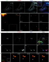
Varp, TI-VAMP and Rab21 co-localization. (A) Varp and TI-VAMP partially localize in HeLa cells co-transfected with RFP–TI-VAMP and Varp–GFP. Images were deconvoluted. Arrows indicate co-localization of Varp and TI-VAMP. Asterisk indicates nucleus. (B) Vesicular co-transport of Varp–GFP and RFP–TI-VAMP (arrowheads) in anterograde and retrograde (data not shown) directions in mouse hippocampal neurons (images from supplementary Movies S1 and S2 online). (C) DIV1 mouse hippocampal neurons were transfected with GFP–Rab21-wt and Varp. Images were deconvoluted. Overlay and inset show co-localization of Varp and TI-VAMP (arrows) or Rab21, Varp and TI-VAMP (arrowheads). The edges of the cellular body and neurites are emphasized by a white dashed line. Asterisk indicates nucleus. (D) Detail of distal axon and growth cone of transfected mouse hippocampal neurons. The probable transition zone of growth cone is emphasized by a white dashed line. (E) RFP–TI-VAMP vesicles were tracked in an axon expressing GFP–Rab21. Vesicular movements of TI-VAMP vesicle are shown in the series of frames corresponding to the boxed region. TI-VAMP-positive vesicles (arrowheads) moved into the peripheral region of the axon, whereas Rab21 was retained in the central region (images from supplementary Movie S3 online). Scale bars, 10 μm. Time in minutes. DIV, days in vitro; GFP, green fluorescent protein; RFP, red fluorescent protein; TI-VAMP, tetanus neurotoxin-insensitive vesicle-associated membrane protein; Varp, Vps9 domain and ankyrin-repeat-containing protein; wt, wild type.
Figure 3
Figure 3
Varp regulates neurite growth in PC12 cells and mouse hippocampal neurons. (A) Rat PC12 cells were treated with siRNAs against scramble, luciferase, Syb2, TI-VAMP or Varp (Varp_r1) for 72 h, differentiated with 100 nM staurosporine for 2–12 h and then immunostained alternatively for TI-VAMP, Syb2 or Varp and tubulin (data not shown). Neurite growth was impaired after the depletion of TI-VAMP and Varp but not of Syb2. (B) The degree of silencing of TI-VAMP, Syb2 and Varp expression in PC12 cells was assessed by western blot analysis. (C) DIV1 mouse hippocampal neurons were co-transfected either with two Varp siRNA oligonucleotides (Varp_m1 and Varp_m2) or scramble siRNA and EGFP as a reporter gene and fixed after a further 72 h. The structure of the longest process, that is, the axon (MAP2 negative) is indicated by arrowheads in the merge. (D,E) Quantification of the effect of Varp silencing on axonal length in GFP-positive cells represented as percentile (D) and average (E). (F) Efficiency of mouse Varp siRNA oligonucleotides assessed by western blotting on mouse L-929 cells. (G) DIV1 mouse hippocampal neurons were transfected with Varp–GFP, GFP–ID or GFP, and stained for GFP (green) and MAP2 (red) after a further 48 h. (H,I) Quantification of the effect of Varp and ID overexpression on axonal length represented as percentile (H) and average (I). Percentile representations in (D,H): values on the x-axis indicate the percentage of axons shorter than the length indicated on the y-axis. The shift towards the bottom indicates decreased axonal growth. See also supplementary information for the mean of axon length. Significance determined by two-tailed unpaired t-test ****P<0.0001, ***P<0.005. Data are shown as mean±s.e.m. Scale bars, 20 μm. DIV, days in vitro; EGFP; enhanced green fluorescent protein; GFP; green fluorescent protein; ID, interaction domain; MAP2, microtubule-associated protein 2; n, number of neurites or axons; siRNA, small interfering RNA; Syb2, synaptobrevin 2; TI-VAMP, tetanus neurotoxin-insensitive vesicle-associated membrane protein; Varp, Vps9 domain and ankyrin-repeat-containing protein.
Figure 4
Figure 4
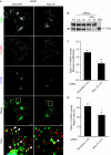
Varp regulates co-localization of TI-VAMP and Rab21. (A) HeLa cells were treated with scramble or Varp (Varp_h2) oligonucleotides for 72 h and then transfected with GFP–Rab21-wt. After 24 h, cells were fixed and labelled for GFP, TI-VAMP and TGN46. Images were processed for deconvolution. Asterisks indicate nuclei. Arrowheads in the inset indicate co-localization of TI-VAMP and Rab21. Scale bar, 10 μm. (B) Western blot showing the efficiency of Varp silencing in HeLa cells transfected with siRNA oligonucleotides against luciferase (Luc), Scramble (Sc) or Varp (Varp_h1 or _h2) compared with not transfected cells. Arrowhead, Varp. (C,D) Co-localization of Rab21 and TI-VAMP in control cells (Scramble) and in Varp-silenced cells represented as Mander's coefficients (C) and as percentage of the number of pixels that overlap between TI-VAMP and Rab21–GFP staining on the total number of pixels for Rab21–GFP staining (D). Significance determined by two-tailed unpaired t-test *P<0.05, **P<0.01. Data are shown as mean±s.e.m. GFP, green fluorescent protein; n, number of cells; NT, not transfected cells; TGN, trans-Golgi network; siRNA, small interfering RNA; TI-VAMP, TI-VAMP, tetanus neurotoxin-insensitive vesicle-associated membrane protein; Varp, Vps9 domain and ankyrin-repeat-containing protein.
Figure 5
Figure 5
Rab21 regulates neurite growth. (A) Effects of Rab21 mutants in differentiating PC12 cells transfected with either GFP–Rab21-wt or -T33N or -Q78L. Arrows indicate neurites and arrowheads indicate small protrusions. Scale bar, 20 μm. (B) Quantification of the neurites longer than 40 μm in differentiated PC12 cells expressing Rab21 mutants. Significance determined by two-tailed unpaired t-test ***P<0.005. Data are shown as mean±s.e.m. (C) Quantification of the length of all neurites (>5 μm) represented as percentile. Significance determined by Kolmogorov test. Rab21 Q78L vs Rab21 wt P<0.05. Rab21 T33N vs Rab21 wt P<0.01. GFP, green fluorescent protein; n, number of neurites; wt, wild type.

References

    1. Alberts P et al. (2003) Cross talk between tetanus neurotoxin-insensitive vesicle-associated membrane protein-mediated transport and L1-mediated adhesion. Mol Biol Cell 14: 4207–4220 - PMC - PubMed
    1. Alberts P, Rudge R, Irinopoulou T, Danglot L, Gauthier-Rouviere C, Galli T (2006) Cdc42 and actin control polarized expression of TI-VAMP vesicles to neuronal growth cones and their fusion with the plasma membrane. Mol Biol Cell 17: 1194–1203 - PMC - PubMed
    1. Boguski MS, McCormick F (1993) Proteins regulating Ras and its relatives. Nature 366: 643–654 - PubMed
    1. Bonifacino JS, Glick BS (2004) The mechanisms of vesicle budding and fusion. Cell 116: 153–166 - PubMed
    1. Chaineau M, Danglot L, Proux-Gillardeaux V, Galli T (2008) Role of HRB in clathrin-dependent endocytosis. J Biol Chem 283: 34365–34373 - PMC - PubMed

Publication types