. 2009 Sep 10;4(9):e6992.
doi: 10.1371/journal.pone.0006992.

Addressing reported pro-apoptotic functions of NF-kappaB: targeted inhibition of canonical NF-kappaB enhances the apoptotic effects of doxorubicin

Affiliations

Addressing reported pro-apoptotic functions of NF-kappaB: targeted inhibition of canonical NF-kappaB enhances the apoptotic effects of doxorubicin

Brian K Bednarski et al. PLoS One. .

Abstract

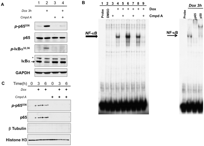
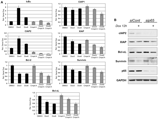
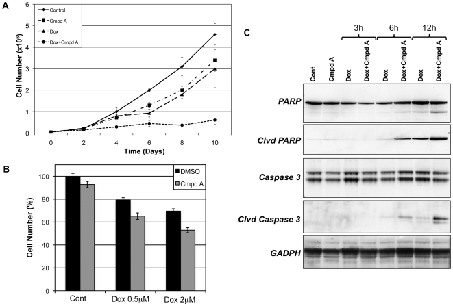
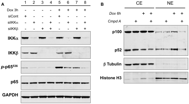
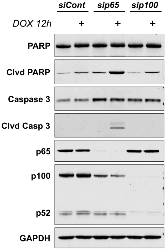
The ability of the transcription factor NF-kappaB to upregulate anti-apoptotic proteins has been linked to the chemoresistance of solid tumors to standard chemotherapy. In contrast, recent studies have proposed that, in response to doxorubicin, NF-kappaB can be pro-apoptotic through repression of anti-apoptotic target genes. However, there is little evidence analyzing the outcome of NF-kappaB inhibition on the cytotoxicity of doxorubicin in studies describing pro-apoptotic NF-kappaB activity. In this study, we further characterize the activation of NF-kappaB in response to doxorubicin and evaluate its role in chemotherapy-induced cell death in sarcoma cells where NF-kappaB is reported to be pro-apoptotic. Doxorubicin treatment in U2OS cells induced canonical NF-kappaB activity as evidenced by increased nuclear accumulation of phosphorylated p65 at serine 536 and increased DNA-binding activity. Co-treatment with a small molecule IKKbeta inhibitor, Compound A, abrogated this response. RT-PCR evaluation of anti-apoptotic gene expression revealed that doxorubicin-induced transcription of cIAP2 was inhibited by Compound A, while doxorubicin-induced repression of other anti-apoptotic genes was unaffected by Compound A or siRNA to p65. Furthermore, the combination of doxorubicin and canonical NF-kappaB inhibition with Compound A or siRNA to p65 resulted in decreased cell viability measured by trypan blue staining and MTS assay and increased apoptosis measured by cleaved poly (ADP-ribose) polymerase and cleaved caspase 3 when compared to doxorubicin alone. Our results demonstrate that doxorubicin-induced canonical NF-kappaB activity associated with phosphorylated p65 is anti-apoptotic in its function and that doxorubicin-induced repression of anti-apoptotic genes occurs independent of p65. Therefore, combination therapies incorporating NF-kappaB inhibitors together with standard chemotherapies remains a viable method to improve the clinical outcomes in patients with advanced stage malignancies.


Conflict of interest statement

Competing Interests: The authors have declared that no competing interests exist.

Figures

Figure 1
Figure 1. Doxorubicin activates canonical NF-κB signaling.
(A) U2OS cells were pretreated with Compound A (Cmpd A, 5 µM) for 1 h and then stimulated with doxorubicin (Dox, 2 µM) for 3 h. Whole cell extracts were evaluated for canonical NF-κB activation by western blot. Doxorubicin induces phosphorylation of IκBα (p-IκBα32,36) and p65 (p-p65536) compared to DMSO-treated controls. Inhibition of the IKK complex with Cmpd A blocks doxorubicin-mediated increase in p-IκBα32,36 and p-p65536. (B) Nuclear extracts were prepared from U2OS cells treated with Dox+/−Cmpd A for 3, 6, and 12 hours and evaluated by EMSA. Doxorubicin treatment results in increased NF-κB DNA-binding activity at all times points. Pretreatment with Cmpd A successfully inhibits the activation of NF-κB by doxorubicin. Supershift analysis confirms that the activated complex of NF-κB contains both p65 and p50 subunits. (C) Western blot of nuclear extracts harvested from cells treated with Dox+/−Cmpd A demonstrates that doxorubicin induces nuclear translocation of p65 phosphorylated at serine 536 (p-p65536), and this is inhibited by the addition of Compound A.
Figure 2
Figure 2. Doxorubicin-mediated repression of anti-apoptotic genes is independent of canonical NF-κB activity.
(A) U2OS cells were stimulated with doxorubicin (Dox) with or without Compound A (Cmpd A) for the indicated times. Total RNA was isolated and evaluated using real time RT-PCR and results are displayed as relative gene expression compared to DMSO-treated controls. Doxorubicin treatment increased transcription of IκBα and cIAP2, decreased transcription of XIAP, Survivin, Bcl-xL, and Bcl2, and did not alter cIAP1 transcription (black bars). Inhibition of NF-κB activation with Cmpd A blocked the induction of IκBα and cIAP2, but had no effect on the doxorubicin-mediated repression of the other anti-apoptotic genes (gray bars). (B) U2OS cells were transfected with either a non-targeting control siRNA (siCont) or siRNA directed against p65 (sip65). After 48 h incubation, the cells were treated with doxorubicin (Dox) for 12 hours and changes in levels of anti-apoptotic proteins were assessed by western blot. Knockdown of the p65 subunit decreased the basal expression of cIAP2, but did not alter baseline protein levels of XIAP, Bcl-xL, or Survivin. Moreover, the absence of p65 did not alter the ability of doxorubicin to repress the expression of these same proteins.
Figure 3
Figure 3. NF-κB inhibition with Compound A enhances the cytotoxicity of doxorubicin through increased apoptosis.
(A) U2OS cells were seeded in 6-well plates and treated every 48 hours with DMSO, doxorubicin (Dox, 5 ng/ml), Compound A (Cmpd A, 5 µM), or Dox+Cmpd A. The total number of live cells was counted at 2-day intervals. While both Dox alone and Cmpd A alone resulted in some decrease in cell growth, the combination of NF-κB inhibition with doxorubicin substantially enhanced the cytotoxic effects of doxorubicin alone. (B) U2OS cells were pretreated with DMSO or Cmpd A and then exposed to Dox at either 0.5 µM or 2 µM for 24 hours. The number of cells was then measured using an MTS assay and displayed as the percent of total cells compared to DMSO-treated controls. Although Cmpd A alone had minimal effect on cell number, its combination with Dox at either dose resulted in decreased number of cells compared to doxorubicin alone. (C) U2OS cells treated with Dox+/−Cmpd A for the indicated time points were evaluated for the presence of the apoptotic markers cleaved PARP and cleaved Caspase 3. Treatment with Dox alone had minimal effects on apoptosis. However, the combination of NF-κB inhibition and Dox resulted in a significant increase in both cleaved PARP and cleaved Caspase 3, which is consistent with increased apoptotic cell death.
Figure 4
Figure 4. Both IKKα and IKKβ contribute to doxorubicin-induced phosphorylation of p65 and doxorubicin induces nuclear accumulation of p52.
(A) U2OS cells were transfected with siRNA constructs targeting IKKα (siIKKα), IKKβ (siIKKβ) or a non-targeting control siRNA (siCont). The cells were then treated with doxorubicin (Dox) for 3 hours. Western blot confirmed that siIKKα and siIKKβ achieved selective knockdown of their respective subunits. Analysis of phosphorylation of p65 (p-p65536) demonstrated that the decrease in either IKKα or IKKβ alone did not completely eliminate the ability of doxorubicin to induce p-p65536. However, knockdown of both catalytic subunits resulted in complete inhibition of the doxorubicin-induced increase in p65 phosphorylation. (B) Cytoplasmic and nuclear extracts were isolated from U2OS cells treated with doxorubicin (Dox) +/− Compound A (Cmpd A) for 6 hours. Immunoblot for p52/p100 confirms an increase in the nuclear translocation of p52 in response to Dox treatment. Inhibition of the IKK complex with Compound A prevented the nuclear accumulation of p52.
Figure 5
Figure 5. Activation of p65, and not p52, is critical to the apoptotic resistance in U2OS cells.
U2OS cells were transfected with siRNA targeting the p65 subunit (sip65), the p100 subunit (sip100), or a non-targeting control (siCont). After 48 hours of incubation with the siRNA, the cells were treated with doxorubicin for 12 hours and the effects on apoptosis were assessed by western blot. Western blot for p65 confirmed significant knockdown. Evaluation of p100 levels revealed a modest decrease of p100 in the cells lacking p65 and complete knockdown in the cells treated with sip100. Importantly, the levels of p52 were not affected by sip65, but were substantially decreased in the absence of its precursor p100. Analysis of the apoptotic markers cleaved PARP and cleaved Caspase 3, revealed a robust increase in apoptosis in response to doxorubicin only in cells lacking the p65 subunit. Cells treated with siCont or sip100 demonstrated no increase in cleaved PARP or cleaved Caspase 3 when treated with doxorubicin.

References

    1. Basseres DS, Baldwin AS. Nuclear factor-kappaB and inhibitor of kappaB kinase pathways in oncogenic initiation and progression. Oncogene. 2006;25:6817–6830. - PubMed
    1. Kim HJ, Hawke N, Baldwin AS. NF-kappaB and IKK as therapeutic targets in cancer. Cell Death Differ. 2006;13:738–747. - PubMed
    1. Shen HM, Tergaonkar V. NFkappaB signaling in carcinogenesis and as a potential molecular target for cancer therapy. Apoptosis. 2009;14:348–363. - PubMed
    1. Wang CY, Mayo MW, Baldwin AS., Jr TNF- and cancer therapy-induced apoptosis: potentiation by inhibition of NF-kappaB. Science. 1996;274:784–787. - PubMed
    1. Wang CY, Guttridge DC, Mayo MW, Baldwin AS., Jr NF-kappaB induces expression of the Bcl-2 homologue A1/Bfl-1 to preferentially suppress chemotherapy-induced apoptosis. Mol Cell Biol. 1999;19:5923–5929. - PMC - PubMed

Publication types

MeSH terms