

Review
doi: 10.1089/ars.2009.2878.
Threading the needle: getting selenocysteine into proteins
Affiliations
- PMID: 19747061
- PMCID: PMC2864665
- DOI: 10.1089/ars.2009.2878
Item in Clipboard
Review
Threading the needle: getting selenocysteine into proteins
Antioxid Redox Signal.
.
Abstract
The co-translational incorporation of selenocysteine (Sec) requires that UGA be recognized as a sense rather than a nonsense codon. This is accomplished by the concerted action of a Sec insertion sequence (SECIS) element, SECIS binding protein 2, and a ternary complex of the Sec specific elongation factor, Sec-tRNA(Sec), and GTP. The mechanism by which they alter the canonical protein synthesis reaction has been elusive. Here we present an overview of the mechanistic perspective on Sec incorporation, highlighting recent advances in the field.
Figures
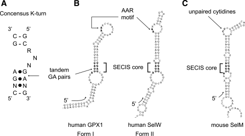
Comparison of K-turn and SECIS elements. (A) The consensus K-turn as described (34). The canonical Watson–Crick base-paired stem is separated from the noncanonical, sheared tandem GA pairs containing, stem. Base pairing marked by dots indicate non-Watson–Crick interactions. (B) Canonical form I and form II SECIS elements are depicted here by the human GPX1 and SelW SECIS elements, respectively. The SECIS core and AAR motifs are in shown in bold and indicated by brackets and arrows. (C) The mouse SelM SECIS element with apical unpaired cytidines is shown for comparison with the canonical SECIS elements in (B). SECIS elements were drawn with the SECISearch program (37) and orientation of the RNAs is indicated by the 5′ arrow at the base of the SECIS elements.
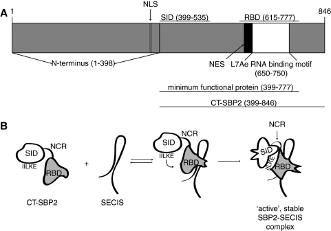
Proposed models for SBP2-SECIS interactions and SBP2 domain structure. (A) Schematic of SBP2 outlining current domain definitions as well as the nuclear localization signal (NLS) and nuclear export signal (NES) described by Papp et al. (56). (B) A cartoon for a model of SBP2 binding the SECIS element. In this model, SBP2 initially interacts with the SECIS via low affinity contacts indicated by the equilibrium arrows. The affinity interaction triggers a conformational change in the RBD that recruits the SID (middle). Subsequent high affinity SECIS interactions and stabilization of the SID-RBD interaction by the residues IILKE526–530 results in an active complex and conformational changes in the SID (right). NCR designates the nonconserved region between the SID and RBD that is not essential for Sec incorporation or SECIS binding (6).
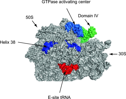
Modeling eEFSec on the ribosome. The cryo-EM model of EF-Tu/Phe-tRNA in the ribosomal pre-accommodated state (PDB coordinates 3EP2) was overlayed onto the high resolution crystal structure of the T. thermophilus 70S ribosome in a post-translocation state containing E and P-site tRNAs and an empty A site (PDB coordinates 1VSP[50S] and 2QNH[30S]). Once the pre-accommodated cryoEM structure was aligned, we then overlaid the crystal structure of GDPNP bound archaeal SelB ( PDB coordinates 1W3B; 38) to derive the final high resolution model shown here. Archaeal SelB is shown in green. 50S and 30S ribosomal subunits are indicated. The GTPase activating center (light blue), Helix 38 of the 50S subunit (dark blue), and E-site tRNA (red) are shown as landmarks. (For interpretation of the references to color in this figure legend, the reader is referred to the web version of this article at www.liebertonline.com/ars ).
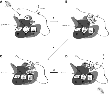
A model for Sec incorporation. In the absence of an SBP2-SECIS complex, eEFSec does not have access to the ribosomal A-site (A). Arrow 1 serves to indicate SECIS binding by SBP2 as described in Fig. 2A. SECIS bound SBP2 is competent to recruit eEFSec (B) which results in a conformational in the SID that signals the ribosome to allow eEFSec to deliver Sec-tRNASec (arrow 2 and C). Arrow 3 indicates GTP hydrolysis by eEFSec and which results in tRNA release and dissociation of eEFSec from the ribosome (D). The fate of SBP2 after Sec incorporation is not known, as noted by the arrow and question mark in D.
References
-
- Alsina B. Corominas M. Berry MJ. Baguña J. Serras F. Disruption of selenoprotein biosynthesis affects cell proliferation in the imaginal discs and brain of Drosophila melanogaster. J Cell Sci. 1999;112:2875–2884. - PubMed
-
- Andersen GR. Pedersen L. Valente L. Chatterjee I. Kinzy TG. Kjeldgaard M. Nyborg J. Structural basis for nucleotide exchange and competition with tRNA in the yeast elongation factor complex eEF1A:eEF1Balpha. Mol Cell. 2000;6:1261–1266. - PubMed
-
- Boulon S. Marmier–Gourrier N. Pradet–Balade B. Wurth L. Verheggen C. Jády BE. Rothé B. Pescia C. Robert MC. Kiss T. Bardoni B. Krol A. Branlant C. Allmang C. Bertrand E. Charpentier B. The Hsp90 chaperone controls the biogenesis of L7Ae RNPs through conserved machinery. J Cell Biol. 2008;180:579–595. - PMC - PubMed
Publication types
MeSH terms
Substances
Grants and funding
LinkOut - more resources
Full Text Sources
Other Literature Sources
