

Thrombospondin-1 (TSP-1) up-regulates tissue inhibitor of metalloproteinase-1 (TIMP-1) production in human tumor cells: exploring the functional significance in tumor cell invasion
- PMID: 19747478
- PMCID: PMC2783349
- DOI: 10.1016/j.yexmp.2009.09.002
Thrombospondin-1 (TSP-1) up-regulates tissue inhibitor of metalloproteinase-1 (TIMP-1) production in human tumor cells: exploring the functional significance in tumor cell invasion
Abstract
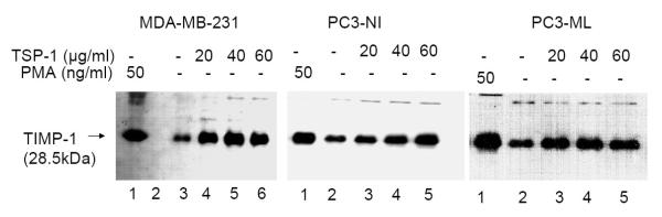
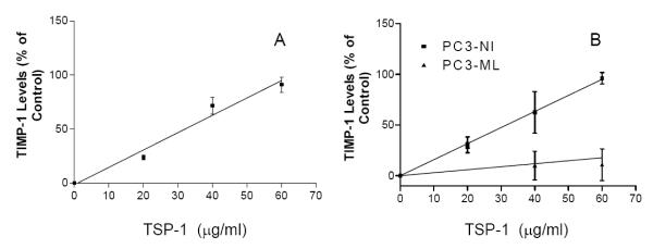
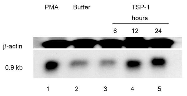
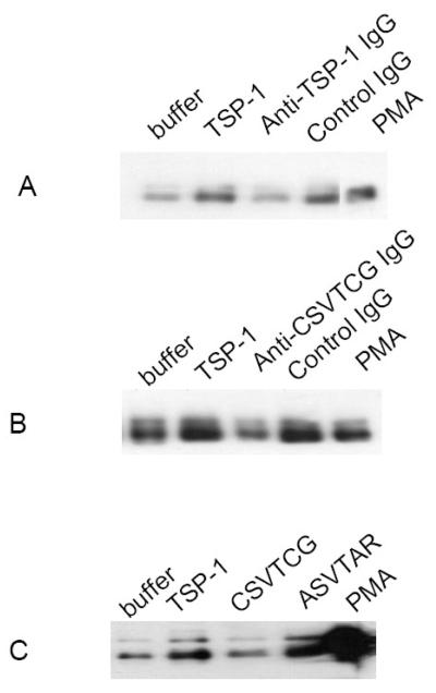
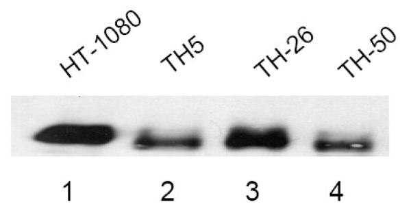
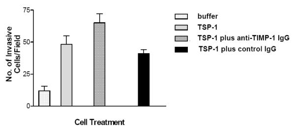
Thrombospondin-1 (TSP-1), a matrix-bound adhesive glycoprotein, has been shown to modulate tumor progression. We previously demonstrated that TSP-1 up-regulates matrix metalloproteinases MMP-2 and MMP-9. Our studies suggested that the balance between MMPs and tissue inhibitors of metalloproteinases (TIMPs) is a key determinant in tumor cell invasion. We now report that TSP-1 up-regulates TIMP-1 expression in both human breast and prostate cancer cell lines. The effect of TSP-1 on TIMP-1 expression was examined in human breast adenocarcinoma cell lines (MDA-MB-231) and human prostate cancer cell lines (PC3-NI and PC3-ML) treated with exogenous TSP-1. TIMP-1 expression was also examined in TSP-1 stably transfected breast cancer cell line (MDA-MB-435). Northern and western blot analysis revealed TIMP-1 mRNA and TIMP-1 protein expression increased with increasing concentrations of TSP-1. This effect was inhibited by antibodies against the type I repeat domain of TSP-1 further suggesting that TSP-1 mediates TIMP-1 secretion. Inhibition of TSP-1 induced TIMP-1 levels increased tumor cell invasion. We conclude that TSP-1 is involved in influencing the critical balance between MMPs and their inhibitors, maintaining the controlled degradation of the extracellular matrix needed to support metastasis and our results may provide an explanation for the divergent activities reported for TSP-1 in tumor progression.
Figures
Similar articles
-
Expression of thrombospondin-1 in human pancreatic adenocarcinomas: role in matrix metalloproteinase-9 production.Pathol Oncol Res. 2001;7(4):251-9. doi: 10.1007/BF03032381. Pathol Oncol Res. 2001. PMID: 11882904
-
Correlation between MMPs and their inhibitors in breast cancer tumor tissue specimens and in cell lines with different metastatic potential.BMC Cancer. 2009 Jan 14;9:20. doi: 10.1186/1471-2407-9-20. BMC Cancer. 2009. PMID: 19144199 Free PMC article.
-
Fibroblasts promote breast cancer cell invasion by upregulating tumor matrix metalloproteinase-9 production.Surgery. 2002 Aug;132(2):220-5. doi: 10.1067/msy.2002.125353. Surgery. 2002. PMID: 12219015
-
Up-regulation of matrix metalloproteinase 9 by thrombospondin 1 in gastric cancer.J Surg Res. 2002 Nov;108(1):51-60. doi: 10.1006/jsre.2002.6452. J Surg Res. 2002. PMID: 12443715
-
The tumor suppressor PTEN inhibits EGF-induced TSP-1 and TIMP-1 expression in FTC-133 thyroid carcinoma cells.Exp Cell Res. 2005 Mar 10;304(1):187-201. doi: 10.1016/j.yexcr.2004.10.026. Epub 2004 Nov 23. Exp Cell Res. 2005. PMID: 15707585
Cited by
-
Down-regulation of TIMP-1 inhibits cell migration, invasion, and metastatic colonization in lung adenocarcinoma.Tumour Biol. 2015 May;36(5):3957-67. doi: 10.1007/s13277-015-3039-5. Epub 2015 Jan 13. Tumour Biol. 2015. PMID: 25578494
-
Cromolyn prevents cerebral vasospasm and dementia by targeting WDR43.Front Aging Neurosci. 2023 Apr 13;15:1132733. doi: 10.3389/fnagi.2023.1132733. eCollection 2023. Front Aging Neurosci. 2023. PMID: 37122373 Free PMC article.
-
Thrombospondins as key regulators of synaptogenesis in the central nervous system.Matrix Biol. 2012 Apr;31(3):170-7. doi: 10.1016/j.matbio.2012.01.004. Epub 2012 Jan 21. Matrix Biol. 2012. PMID: 22285841 Free PMC article. Review.
-
THBS1 regulates trophoblast fusion through a CD36-dependent inhibition of cAMP, and its upregulation participates in preeclampsia.Genes Dis. 2020 Jun 8;8(3):353-363. doi: 10.1016/j.gendis.2020.05.007. eCollection 2021 May. Genes Dis. 2020. PMID: 33997182 Free PMC article.
-
Structural insights into regulation of CCN protein activities and functions.J Cell Commun Signal. 2023 Jun;17(2):371-390. doi: 10.1007/s12079-023-00768-5. Epub 2023 May 28. J Cell Commun Signal. 2023. PMID: 37245184 Free PMC article. Review.
References
-
- Albo D, Tuszynski GP. Thrombospondin-1 up-regulates tumor cell invasion through the urokinase plasminogen activator receptor in head and neck cancer cells. J Surg Res. 2004;120:21–6. - PubMed
-
- Chambers AF. MDA-MB-435 and M14 cell lines: identical but not M14 melanoma? Cancer Res. 2009;69:5292–3. - PubMed
-
- Haas TL, et al. Three-dimensional type I collagen lattices induce coordinate expression of matrix metalloproteinases MT1-MMP and MMP-2 in microvascular endothelial cells. J Biol Chem. 1998;273:3604–10. - PubMed
-
- Lawler J. The structural and functional properties of thrombospondin. Blood. 1986;67:1197–209. - PubMed
Publication types
MeSH terms
Substances
Grants and funding
LinkOut - more resources
Full Text Sources
Medical
Research Materials
Miscellaneous
