. 2009 Nov 2;8(11):1283-9.
doi: 10.1016/j.dnarep.2009.08.002. Epub 2009 Sep 10.

Structural basis for the lack of opposite base specificity of Clostridium acetobutylicum 8-oxoguanine DNA glycosylase

Affiliations

Structural basis for the lack of opposite base specificity of Clostridium acetobutylicum 8-oxoguanine DNA glycosylase

Frédérick Faucher et al. DNA Repair (Amst). .

Abstract

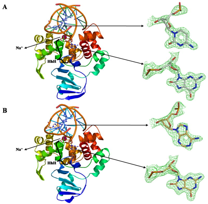
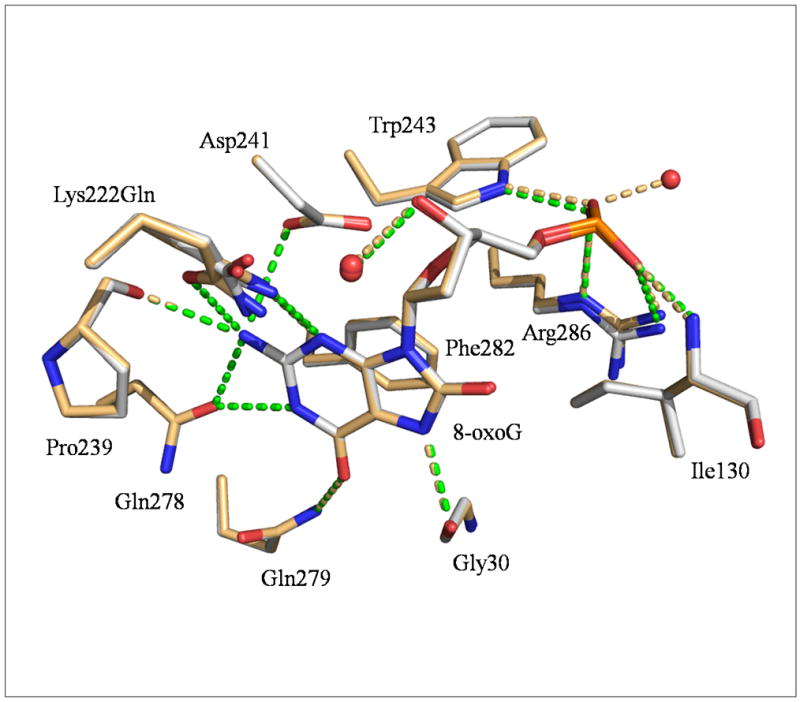
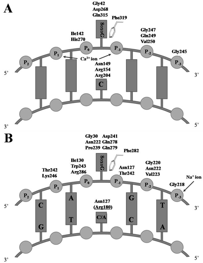
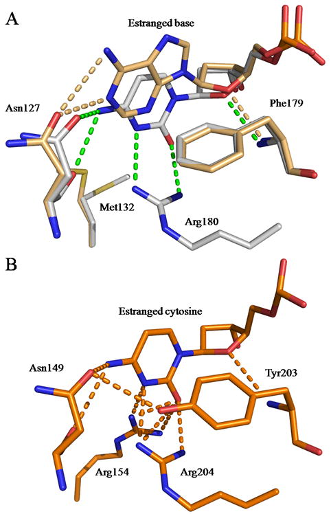
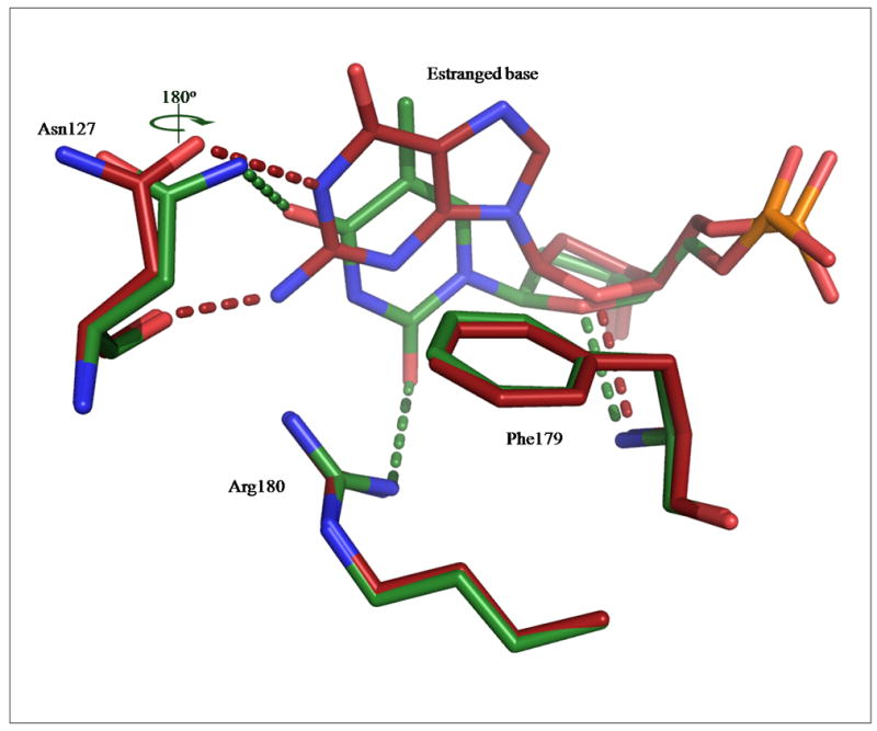
7,8-Dihydro-8-oxoguanine (8-oxoG) is the major oxidative product of guanine and the most prevalent base lesion observed in DNA molecules. Because 8-oxoG has the capability to form a Hoogsteen pair with adenine (8-oxoG:A) in addition to a normal Watson-Crick pair with cytosine (8-oxoG:C), this lesion can lead to a G:C-->T:A transversion after replication. However, 8-oxoG is recognized and excised by the 8-oxoguanine DNA glycosylase (Ogg) of the base excision repair pathway. Members of the Ogg1 family usually display a strong preference for a C opposite the lesion. In contrast, the atypical Ogg1 from Clostridium actetobutylicum (CacOgg) can excise 8-oxoG when paired with either one of the four bases, albeit with a preference for C and A. Here we describe the first high-resolution crystal structures of CacOgg in complex with duplex DNA containing the 8-oxoG lesion paired to cytosine and to adenine. A structural comparison with human OGG1 provides a rationale for the lack of opposite base specificity displayed by the bacterial Ogg.


Conflict of interest statement

Conflict of interest statement

The authors declare that there are no conflicts of interest.

Figures

Figure 1
Figure 1. Overall fold of CacOggK222Q in complex with DNA containing A) 8-oxoG:C and B) 8-oxoG:A
Ribbon diagrams of CacOggK222Q in complex with DNA containing A) 8-oxoG:C and B) 8-oxoG:A Proteins are colored according to the amino acid sequence going from cold blue to warm red from N- to C-terminal. A simulated annealing omit map (green) contoured at 3σ is shown for each of the estranged bases and 8-oxoG. The sodium atom is colored in pink in both panels and the HhH motif is labeled. All figures were prepared using PYMOL [38].
Figure 2
Figure 2. Close-up view of CacOgg residues interacting with 8-oxoG
CacOggK222Q/8-oxoG:C and/8-oxoG:A are superimposed. Only residues involved in H-bonds and stacking interactions with 8-oxoG are depicted. The CacOggK222Q/8-oxoG:C carbon atoms are shown in light grey and those of the 8-oxoG:A complex in light orange. Gln222 appears to adopt alternate conformations in both complexes. H-bonds are represented by green dashed lines for 8-oxoG:C complex and light orange dash line for 8-oxoG:A complex. The H-bond between 8-oxoG N7 H atom and the main chain carbonyl of Gly30 has been shown to be essential for the recognition of 8-oxoG in hOGG1[26, 39].
Figure 3
Figure 3. Schematic representation of Ogg1-DNA interactions
Schematic representation of A) DNA interactions with hOGG1 (PDB ID code 1EBM [26]) and B) interactions of DNA with CacOggK222Q/8-oxoG:C/A complexes. hOGG1-Phe319 and CacOgg-Phe282 stack against the 6-membered ring of 8-oxoG. Arg180 (in parenthesis and underlined) in panel B makes a H-bond only when the estranged base is a cytosine. Metal ion interactions are depicted.
Figure 4
Figure 4. CacOgg interactions with the estranged nucleoside
A) CacOggK222Q/8-oxoG:C and/8-oxoG:A are superimposed. Only residues involved in H-bonds and stacking interactions with the estranged base are depicted. CacOggK222Q/8-oxoG:C carbon atoms are shown in light grey and those in the 8-oxoG:A complex are colored in light orange. Phe179 is involved in both hydrophobic and hydrophilic interactions. B) Interactions made by hOGG1 (PDB ID code 1EBM) [26] with estranged cytosine. Tyr203 is involved in both hydrophobic and hydrophilic interactions and helps stabilize the side chain of Asn149. H-bonds are represented by green dashed lines for CacOgg/8-oxoG:C or by the color corresponding to their structure.
Figure 5
Figure 5. Putative interactions of guanine and thymine with CacOgg at the estranged base binding site
The figure shows the putative interactions that CacOgg may make with G (red) or T (green) as the estranged base. The putative H-bonds are depicted in the same color as their corresponding model. Asn127 most likely performs a 180° rotation around its Cγ to interact with the O4 acceptor of thymine. In both models, the estranged base stacks against of Phe179. The hydrophilic interactions are extrapolated from the CacOgg/8-oxoG:C and/8-oxoG:A complexes.

References

    1. Fraga CG, Shigenaga MK, Park JW, Degan P, Ames BN. Oxidative damage to DNA during aging: 8-hydroxy-2′-deoxyguanosine in rat organ DNA and urine. Proc Natl Acad Sci U S A. 1990;87:4533–4537. - PMC - PubMed
    1. Kasai H, Crain PF, Kuchino Y, Nishimura S, Ootsuyama A, Tanooka H. Formation of 8-hydroxyguanine moiety in cellular DNA by agents producing oxygen radicals and evidence for its repair. Carcinogenesis. 1986;7:1849–1851. - PubMed
    1. Grollman AP, Moriya M. Mutagenesis by 8-oxoguanine: an enemy within. Trends Genet. 1993;9:246–249. - PubMed
    1. Shibutani S, Takeshita M, Grollman AP. Insertion of specific bases during DNA synthesis past the oxidation-damaged base 8-oxodG. Nature. 1991;349:431–434. - PubMed
    1. Kuchino Y, Mori F, Kasai H, Inoue H, Iwai S, Miura K, Ohtsuka E, Nishimura S. Misreading of DNA templates containing 8-hydroxydeoxyguanosine at the modified base and at adjacent residues. Nature. 1987;327:77–79. - PubMed

Publication types