. 2009 Sep 9;17(9):1205-12.
doi: 10.1016/j.str.2009.08.002.

Secondary structure of Huntingtin amino-terminal region

Affiliations

Secondary structure of Huntingtin amino-terminal region

Mee Whi Kim et al. Structure. .

Abstract

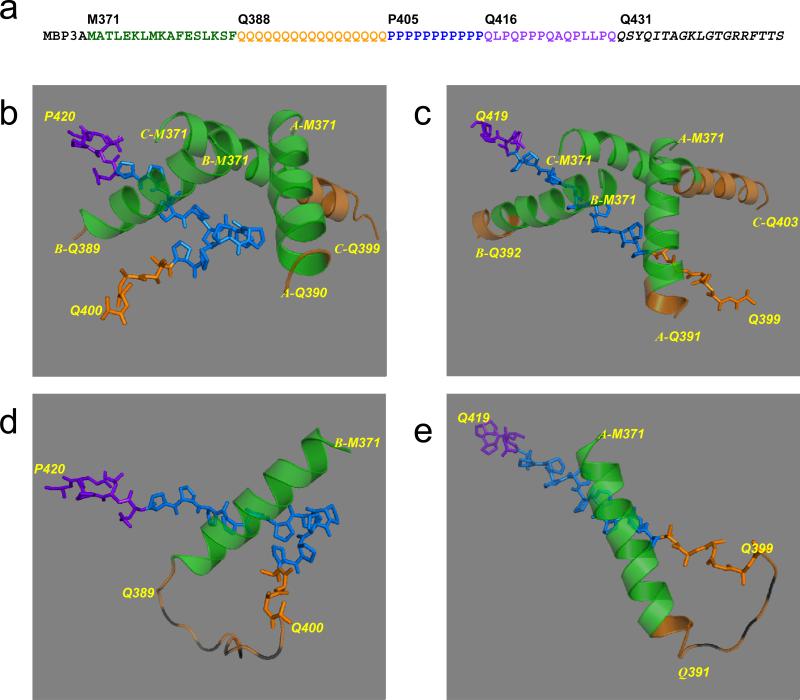
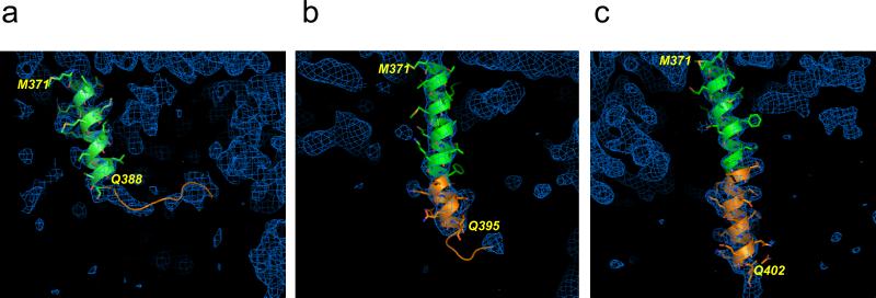
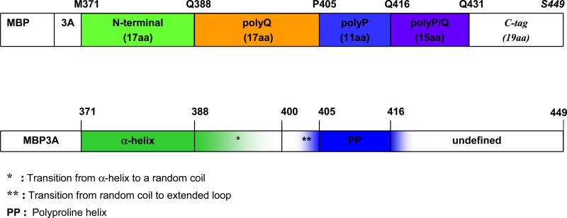
Huntington's disease is a genetic neurodegenerative disorder resulting from polyglutamine (polyQ) expansion (>36Q) within the first exon of Huntingtin (Htt) protein. We applied X-ray crystallography to determine the secondary structure of the first exon (EX1) of Htt17Q. The structure of Htt17Q-EX1 consists of an amino-terminal alpha helix, poly17Q region, and polyproline helix formed by the proline-rich region. The poly17Q region adopts multiple conformations in the structure, including alpha helix, random coil, and extended loop. The conformation of the poly17Q region is influenced by the conformation of neighboring protein regions, demonstrating the importance of the native protein context. We propose that the conformational flexibility of the polyQ region observed in our structure is a common characteristic of many amyloidogenic proteins. We further propose that the pathogenic polyQ expansion in the Htt protein increases the length of the random coil, which promotes aggregation and facilitates abnormal interactions with other proteins in cells.


Figures

Fig. 1
Fig. 1. Secondary structure of Htt17Q-EX1
(a) Amino acid sequence of MBP-Htt17Q-EX1. MBP3A denotes the maltose binding protein followed by a 3Ala linker. M371 to G429 is the sequence of Htt17Q-EX1, which is subdivided into 17 aa N-terminal region (M371 to F387), poly17Q region (Q388 to Q404), poly11P region (P405 to P415), and 15 aa mixed P/Q region (Q416 to Q430). The sequence from Q431 to the C-terminus is the 19 aa tag added to facilitate crystallization. (b) The structure of Htt17Q-EX1 trimer from c95 crystal. The structures of MBP and 3A linker are removed for clarity. The amino-terminal α-helix of Htt17Q-EX1 extends from Met371 to Phe387 (Green). The following poly17Q region (Orange) is α-helical and unstructured (random coil). The poly11P region (Blue) adopts PP-helix (shown as stick model) in the “kinked” conformation. The initial part of polyP/Q region (Purple) is also in PP-helix conformation (shown as stick model). The terminal part of poly17Q region (Orange) is in the extended conformation (shown as stick model) (c) The structure of Htt17Q-EX1 trimer from cHg99 crystal. Same as on panel b but the Poly11P region is in the “straight” conformation. (d) The complete structure of B molecule of Htt17Q-EX1 monomer from c95 crystal. The striped orange loops are for the random coil region between Gln389 and Gln399, which is invisible on the map. (e) The complete structure of A molecule of Htt17Q-EX1 monomer from cHg99 crystal. The striped orange loops are for the random coil region between Gln391 and Gln398, which is invisible on the map.
Fig. 2
Fig. 2. Structure of amino-terminal and poly17Q regions of Htt17Q-EX1
The structures of amino-terminal region (Green) and poly17Q region (Orange) of Htt17Q-EX1 are shown for three different molecules with variable length of poly17Q α-helix. Also shown are corresponding regions of electron density maps contoured at 1.0σ (Blue). (a) The short poly17Q helix makes transition to loop at Gln388 (molecule A, crystal c95). (b) The medium poly17Q helix makes transition to loop at Gln395 (molecule C, crystal c90). (c) The long poly17Q helix extends for the length of polyQ region until Gln402 (molecule C, crystal cHg99).
Fig. 3
Fig. 3. Schematic diagram of the secondary structure elements of Htt17Q-EX1
The first bar represents the sequence of MBP-Htt17Q-EX1 construct which consists of MBP protein (not shown to scale), 3 Alanines linker (3A), 17 aa long N-terminal region of Htt (green), polyQ region with 17 glutamine residues (poly17Q, orange), polyP region with 11 proline residues (poly11P, blue)), 15 aa long mixed polyglutamine and polyproline region (polyP/Q, purple), and C-terminal tag of 19 aa (not coloured). The second bar is a summary of structural information obtained from analysis of Htt17Q-EX1 structures resolved in the seven crystals. The main secondary structure elements of Htt17Q-EX1 are shown: α-helix (bold green), transition from α-helix to random coil region (shaded green), random coil (uncoloured), transition to extended loop region (shaded blue), PP helix (bold blue), transition from PP helix to unstructured region (shaded blue).

Comment in

References

    1. Altschuler EL, Hud NV, Mazrimas JA, Rupp B. Random coil conformation for extended polyglutamine stretches in aqueous soluble monomeric peptides. J Pept Res. 1997;50:73–75. - PubMed
    1. Armen RS, Bernard BM, Day R, Alonso DO, Daggett V. Characterization of a possible amyloidogenic precursor in glutamine-repeat neurodegenerative diseases. Proc Natl Acad Sci U S A. 2005;102:13433–13438. - PMC - PubMed
    1. Atwal RS, Xia J, Pinchev D, Taylor J, Epand RM, Truant R. Huntingtin has a membrane association signal that can modulate huntingtin aggregation, nuclear entry and toxicity. Hum Mol Genet. 2007;16:2600–2615. - PubMed
    1. Bennett MJ, Huey-Tubman KE, Herr AB, West AP, Jr., Ross SA, Bjorkman PJ. Inaugural Article: A linear lattice model for polyglutamine in CAG-expansion diseases. Proc Natl Acad Sci U S A. 2002;99:11634–11639. - PMC - PubMed
    1. Bevivino AE, Loll PJ. An expanded glutamine repeat destabilizes native ataxin-3 structure and mediates formation of parallel beta -fibrils. Proc Natl Acad Sci U S A. 2001;98:11955–11960. - PMC - PubMed

Publication types

LinkOut - more resources