. 2009 Sep 11;35(5):642-56.
doi: 10.1016/j.molcel.2009.07.002.

The interaction of NSBP1/HMGN5 with nucleosomes in euchromatin counteracts linker histone-mediated chromatin compaction and modulates transcription

Affiliations

The interaction of NSBP1/HMGN5 with nucleosomes in euchromatin counteracts linker histone-mediated chromatin compaction and modulates transcription

Mark Rochman et al. Mol Cell. .

Abstract

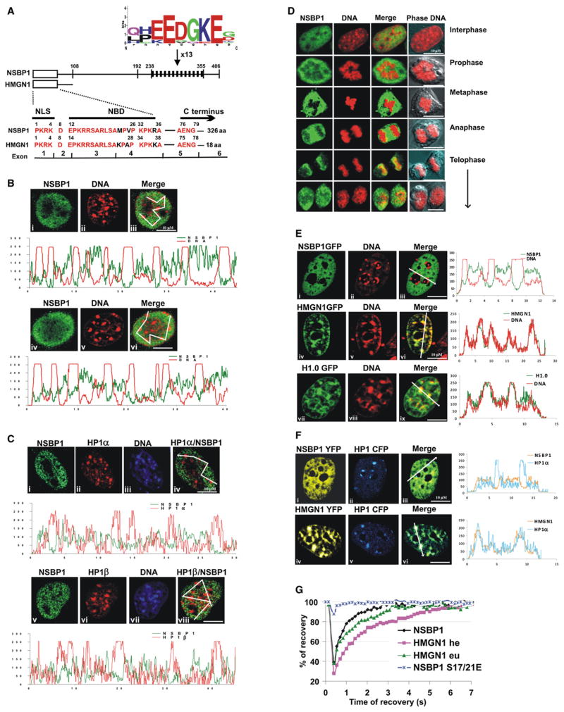
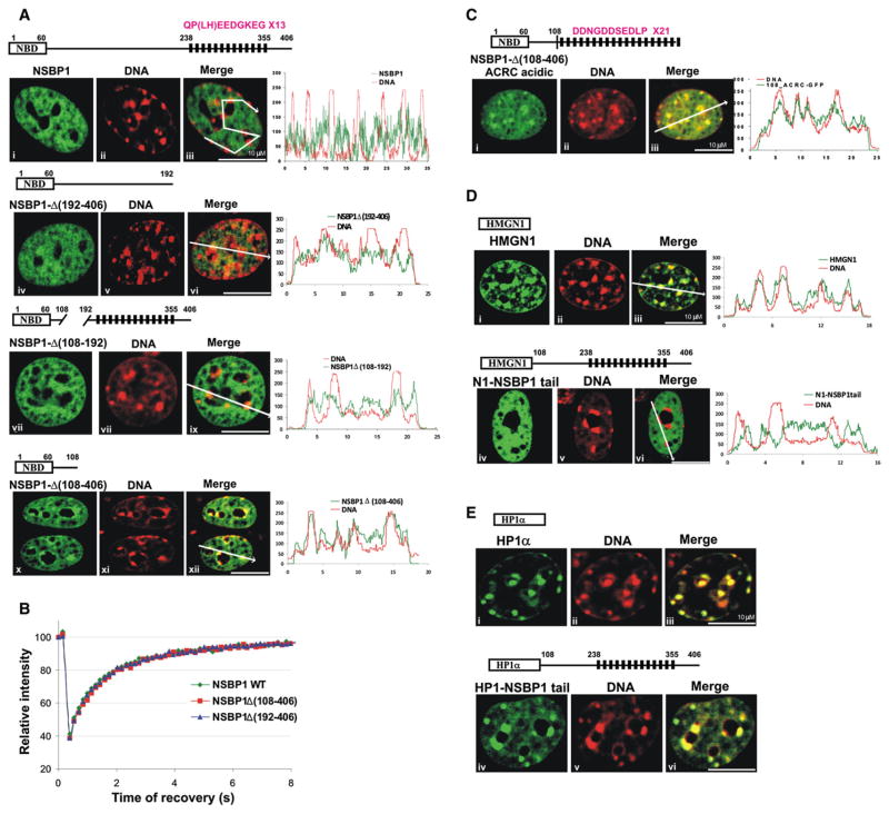
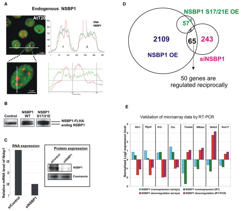
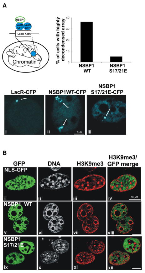
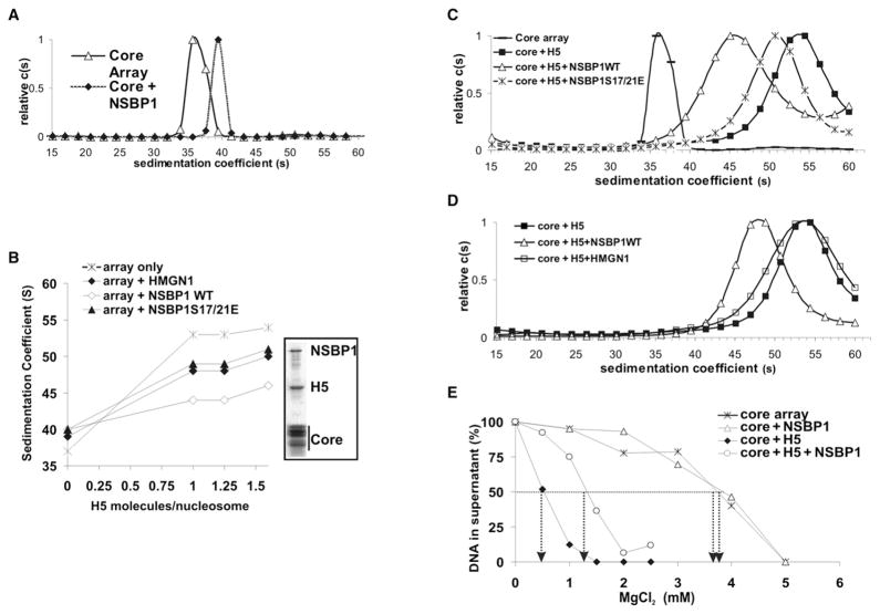
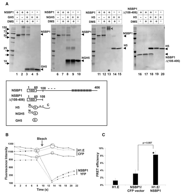
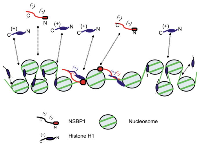
Structural changes in specific chromatin domains are essential to the orderly progression of numerous nuclear processes, including transcription. We report that the nuclear protein NSBP1 (HMGN5), a recently discovered member of the HMGN nucleosome-binding protein family, is specifically targeted by its C-terminal domain to nucleosomes in euchromatin. We find that the interaction of NSBP1 with nucleosomes alters the compaction of cellular chromatin and that in living cells, NSBP1 interacts with linker histones. We demonstrate that the negatively charged C-terminal domain of NSBP1 interacts with the positively charged C-terminal domain of H5 and that NSBP1 counteracts the linker histone-mediated compaction of a nucleosomal array. Dysregulation of the cellular levels of NSBP1 alters the transcription level of numerous genes. We suggest that mouse NSBP1 is an architectural protein that binds preferentially to euchromatin and modulates the fidelity of the cellular transcription profile by counteracting the chromatin-condensing activity of linker histones.


Figures

Figure 1
Figure 1. NSBP1 Protein Localizes to Nucleosomes in Euchromatin
(A) Sequence similarity between HMGN1 and the N-terminal region of NSBP1. The schematic structure of NSBP1 and HMGN1 is shown above the sequence. The position of the exons is indicated below the sequence. NLS, nuclear localization signal; NBD, nucleosome binding domain. The location of the negatively charged sequence repeat is indicated by black bars; the consensus sequence of the repeat is shown above the diagram. (B) NSBP1 does not localize to constitutive heterochromatin. Endogenous NSBP1 protein is visualized by immunofluorescence in green (i and iv) and DNA by Hoechst (ii and v). The merged images (iii and vi) and their localization profiles indicate that NSBP1 does not colocalize with constitutive heterochromatin (compact dots of DNA). (C) NSBP1 does not colocalize with HP1 proteins. Immunostaining of endogenous NSBP1 protein is shown in green (i and v), HP1α (ii) or HP1β (vi) is shown in red, and corresponding DNA staining in blue (iii and vii). Merged images of NSBP1 and HP1 proteins (iv and viii) and their localization profiles are shown. Lack of yellow color indicates that NSBP1 does not colocalize with HP1α or HP1β. (D) Endogenous NSBP1 (green) does not colocalize with metaphase chromosome DNA (red) throughout the mitosis. (E) In live cells, NSBP1-GFP (i) is excluded from constitutive heterochromatin (ii, red), as shown by the absence of yellow (iii) and by the localization profiles. Controls of live cells expressing either HMGN1-GFP (iv) or H1-GFP (vii) indicate that these two proteins localize to heterochromatin. (F) YFP-fused NSBP1 (i) or YFP-HMGN1 (iv) were cotransfected with CFP-fused HP1α (ii and v) and visualized in living cells. Merge images and colocalization profiles of NSBP1, HMGN1, and HP1α are shown. NSBP1 does not while HMGN1 does colocalize with HP1α in living cells. (G) FRAP recovery curves for NSBP1-GFP and for HMGN1-GFP in either euchromatin (eu) or heterochromatin (he) indicate that, in living cells, both bind to chromatin. The extremely fast recovery of the FRAP signal for the NSBP1S17/21E indicates that this mutant does not bind to chromatin.
Figure 2
Figure 2. NSBP1 Is Specifically Targeted to Euchromatin by Its C-Terminal Tail
(A) Intranuclear location of either NSBP1 protein (a–c) or various C-terminal deletion mutants (iv–vi, vii–ix, and x–xii), all expressed as GFP fusion proteins, compared to that of DNA (red), in living cells. A schematic diagram of the expressed proteins is shown above the corresponding images. Merge images (iii, vi, ix, and xii) and colocalization profiles demonstrate that progressive deletion of the C-terminal domain leads to a progressive relocation of the protein to heterochromatin. (B) FRAP analyses of NSBP1-GFP and its C-terminal deletion mutants indicate the C-terminal tail does not affect the affinity of the protein to nucleosomes. (C) A chimeric protein in which the C-terminal domain of NSBP1 protein was exchanged with the acidic C-terminal domain of human ACRC protein localized to heterochromatin. Schematic presentation of acidic portion of ACRC protein fused to tailless NSBP1 is shown above the images. Black rectangles represent the 21 repeats in ACRC. Consensus sequence of the repeat is shown (compare to Figure 2A). (D) The NSBP1 C-terminal domain targets HMGN1 to euchromatin. Shown are the location of wild-type HMGN1 (i–iii) and the HMGN1 fused to the NSBP1 tail (iv–vi) in live cells. The merged images and the localization profile demonstrate that the wild-type HMGN1, but not the HMGN1-NSBP1 chimera, colocalizes with heterochromatin (DNA). (E) The C-terminal domain of NSBP1 does not affect the chromatin interactions of HP1α. Wild-type HP1α (i) and HP1α fused to NSBP1 tail (iv–vi) were visualized in live cells. Colocalization of the proteins with Hoechst staining of DNA (ii and v) showed enrichment of both wild-type HP1α (iii) and HP1α fused to NSBP1 tail (v) in heterochromatin, as judged by the appearance of yellow color in merge images (iii and vi).
Figure 3
Figure 3. NSBP1 Modulates the Transcription Profile of AtT20 Cells
(A) Fluorescent images of mouse pituitary AtT20 cells demonstrate that NSBP1 is depleted from constitutive heterochromatin. Localization profiles are shown for two cells, labeled 1 and 2, and a magnification of cell 2. (B) Western blot analysis of AtT20 cells stably transfected with vectors expressing FLAG-HA (FLHA)-tagged wild-type (WT) NSBP1 or NSBP1S17/21E. Note that the levels of the exogenous protein are similar to the endogenous NSBP1 (endog NSBP1). (C) Downregulation of NSBP1 expression by siRNA. Shown are RT-PCR and western blot analysis of AtT20 cells treated with siRNA against NSBP1 or control siRNA. (D) Venn diagram depicting the overlap of genes whose expression is altered by either overexpressing wild-type NSBP1 (NSBP1 OE) or the NSBP1S17/21E mutant (NSBP1S17/21E OE) or by siRNA-mediated downregulation of NSBP1 (siNSBP1). (E) Validation of microarray data. Shown is normalized log 2 expression of eight selected genes whose expression was reciprocally up- and downregulated by the overexpression or deletion of NSBP1 in AtT20 cells.
Figure 4
Figure 4. NSBP1 Induces Large-Scale Chromatin Decondensation
(A) The interaction of NSBP1 with the nucleosomes decondenses a LacO chromatin array. Schematic drawing (top left) demonstrates the strategy for tethering NSBP1 protein to the array by fusion to LacR-CFP. The condensed array, visualized by tethering LacR-CFP (i), appears as a compact dot. Tethering of the wild-type NSBP1-CFP (ii) to the array induced formation of highly decondensed array, as judged by formation of extended structures with irregular shape. In contrast, tethering of NSBP1S17/21E mutant, which does not bind to nucleosomes (iii), did not unfold the condensed chromatin array. Bar graph above the images indicates the percent of cells containing highly decondensed arrays following tethering of either wild-type or NSBP1S17/21E mutant to the arrays. Thirty cells of each sample were examined. (B) NSBP1 expression alters the global organization of chromatin. The global organization of the constitutive heterochromatin is visualized by DNA staining with Hoechst (ii, vi, and x) or H3K9me3 staining (iii, vii, and xi). Expression of NSBP1 leads to disappearance of the dense heterochromatic foci and condensation of heterochromatin around the nucleoli (vii). Merge images of GFP and H3K9me3 (iv, viii, and xii) demonstrate a lack of colocalization of NSBP1 protein with constitutive heterochromatin.
Figure 5
Figure 5. NSBP1 Counteracts the Linker-Histone-Dependent Chromatin Compaction
(A) NSBP1 does not decrease the compaction of a nucleosome array in the absence of linker histone H5. Shown are the sedimentation profiles of core arrays and core arrays to which NSBP1 was added at a molar ratio of ~2 NSBP1 molecules per core particle (CP). (B) Efficient prevention of H5-mediated chromatin compaction by NSBP1. Shown are sedimentation coefficients of nucleosome arrays incubated with increasing amounts of H5 in the presence of the indicated proteins. NSBP1 and HMGN1 were added at a ratio of two molecules per nucleosome. The Coomassie-stained picture of the SDS-PAGE gel in the boxed area shows that both histone H5 and NSBP1 are bound to the assembled array. (C) NSBP1, but not the NSBP1S17/S21E mutant, unfolds H5-containing arrays. NSBP1 or NSBP1S17/21E proteins were added to a folded array already containing H5 at a molar ratio of ~2 NSBP1 molecules per CP. (D) HMGN1 does not unfold H5-containing arrays. To account for relative protein levels in the cell, 2 HMGN1 but only 0.57 NSBP1 molecules per each H5-containing CP were added. (E) NSBP1 specifically counteracts H5-dependent self-association of the chromatin arrays. Self-association assays were performed with increased concentrations of magnesium chloride and measured as described in the Experimental Procedures. +H5 arrays contained two molecules of histone H5 per CP; +NSBP1 arrays contained two molecules NSBP1 per CP. Arrows indicate magnesium chloride concentration required for 50% of arrays to precipitate. In all samples, the concentration of the core arrays (12 × 207 repeats, see Experimental Procedures) was 25 ng/μl (183 nM mononucleosomes).
Figure 6
Figure 6. NSBP1 Interacts Directly with Linker Histone H5 Both In Vitro and In Vivo
(A) In vitro crosslinking experiments. Full-length NSBP1 and histone H5 or their deletion mutants (see the schematic representation on the bottom of panel A; in this panel, NBD is the nucleosomal NBD of NSBP1, and G is the globular region of H5) were crosslinked by Dimethyl suberimidate (DMS) and analyzed by 15% SDS-PAGE. Lanes 2, 4, 7, 9, 12, 14, 17, and 19 represent the proteins crosslinked individually, whereas lanes 3, 8, 13, and 18 shows the products of crosslinking between H5 and NSBP1 proteins or various deletion mutants. Arrow in line 13 indicates the specific NSBP1-H5 complex formed after crosslinking. Note the disappearance of monomers of NSBP1 and H5 only in lane 13. (B) Detection of linker histone:NSBP1 interaction in living cells by fluorescence resonance energy transfer (FRET). Mouse NIH 3T3 were cotransfected with plasmids expressing H1.E-CFP and NSBP1-YFP proteins. Acceptor photobleaching FRET was performed as described in the Experimental Procedures. Shown is a graphic presentation of fluorescence intensity in three representative ROIs following the bleach of the NSBP1-YFP acceptor. Note the increased fluorescence intensity of H1.E-CFP donor (arrow). (C) Summary of FRET efficiency. The H1.E bar alone is a control for false FRET; NSBP1/CFP vector bar measures random FRET; H1.E/NSBP1 measures the interactions between H1.E-CFP donor and NSBP1-YFP acceptor. Error bars represent standard error (n = 15). P value was calculated using Student’s t test. Interaction between H1.E and NSBP1 was found to be highly statistically significant (*).
Figure 7
Figure 7. Model of the Effect of NSBP1 on Chromatin Compaction
In living cells, H1 and NSBP1 bind dynamically to chromatin (dotted lines). The binding of NSBP1 to H1-containing nucleosomes juxtaposes the C-terminal domains of the proteins and counteracts the H1-induced stabilization of a compact chromatin structure.

References

    1. Alfonso PJ, Crippa MP, Hayes JJ, Bustin M. The footprint of chromosomal proteins HMG-14 and HMG-17 on chromatin subunits. J Mol Biol. 1994;236:189–198. - PubMed
    1. Bianchi ME, Agresti A. HMG proteins: dynamic players in gene regulation and differentiation. Curr Opin Genet Dev. 2005;15:496–506. - PubMed
    1. Brown DT, Izard T, Misteli T. Mapping the interaction surface of linker histone H1(0) with the nucleosome of native chromatin in vivo. Nat Struct Mol Biol. 2006;13:250–255. - PMC - PubMed
    1. Bustin M. Regulation of DNA-dependent activities by the functional motifs of the high-mobility-group chromosomal proteins. Mol Cell Biol. 1999;19:5237–5246. - PMC - PubMed
    1. Bustin M. Chromatin unfolding and activation by HMGN(*) chromosomal proteins. Trends Biochem Sci. 2001;26:431–437. - PubMed

Publication types

MeSH terms

Associated data