. 2009 Sep 11;35(5):669-82.
doi: 10.1016/j.molcel.2009.07.013.

Structure of the Siz/PIAS SUMO E3 ligase Siz1 and determinants required for SUMO modification of PCNA

Affiliations

Structure of the Siz/PIAS SUMO E3 ligase Siz1 and determinants required for SUMO modification of PCNA

Ali A Yunus et al. Mol Cell. .

Abstract

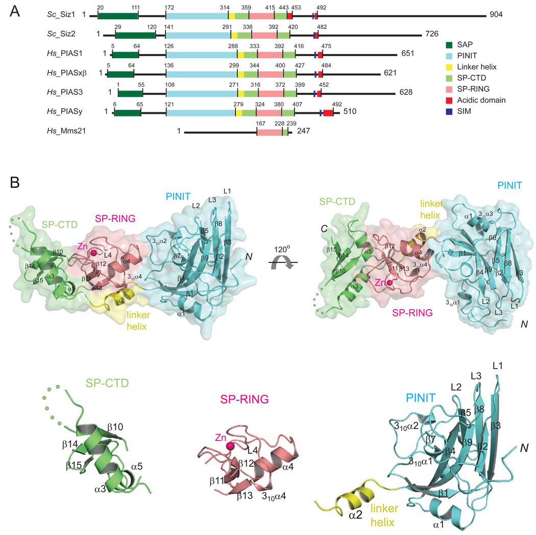
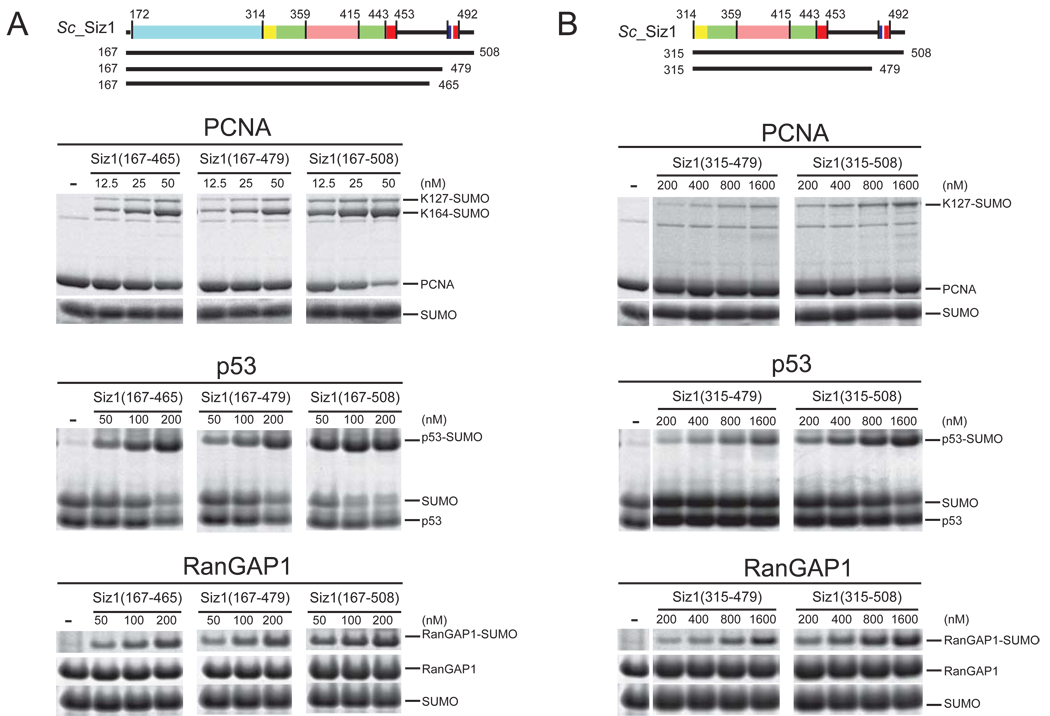
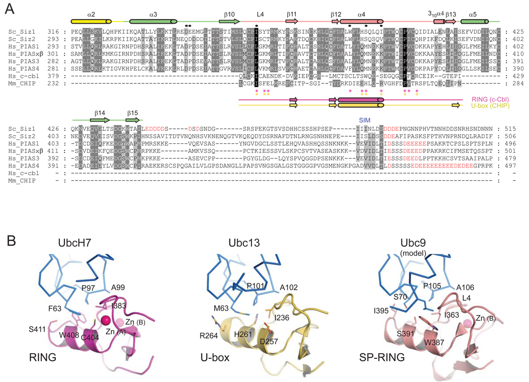
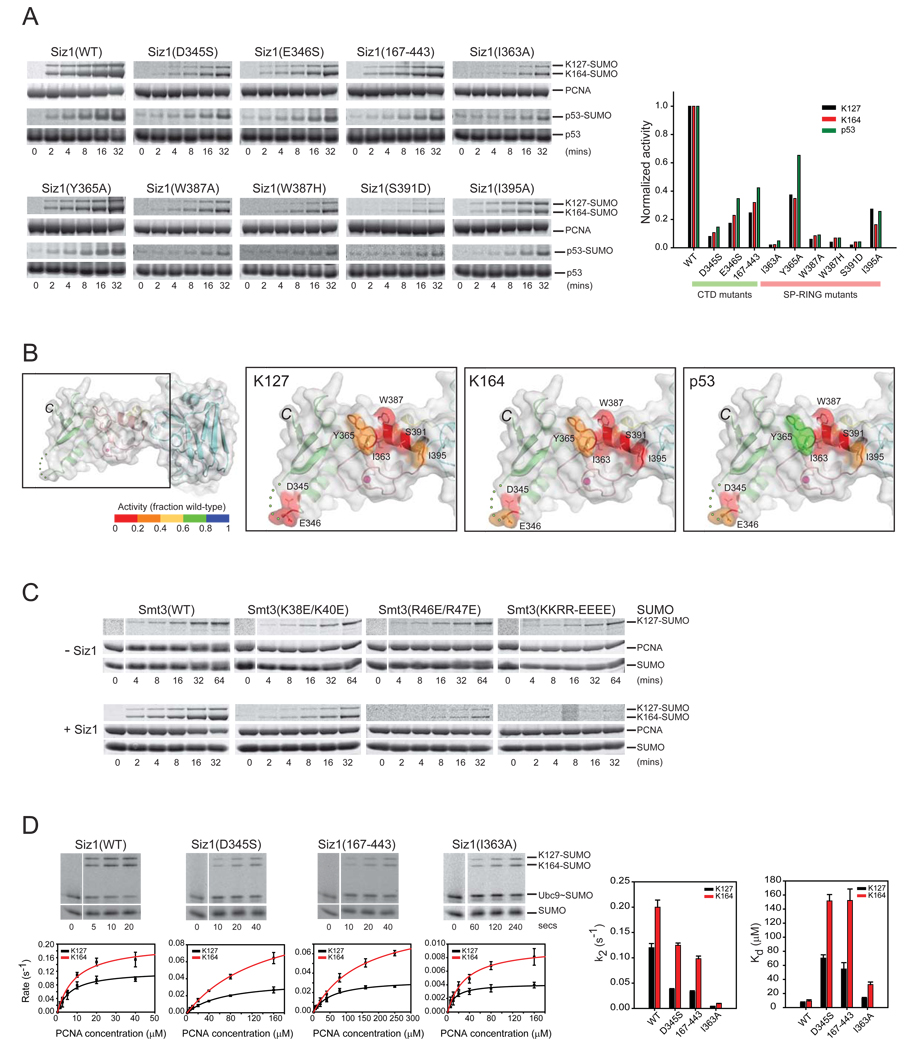
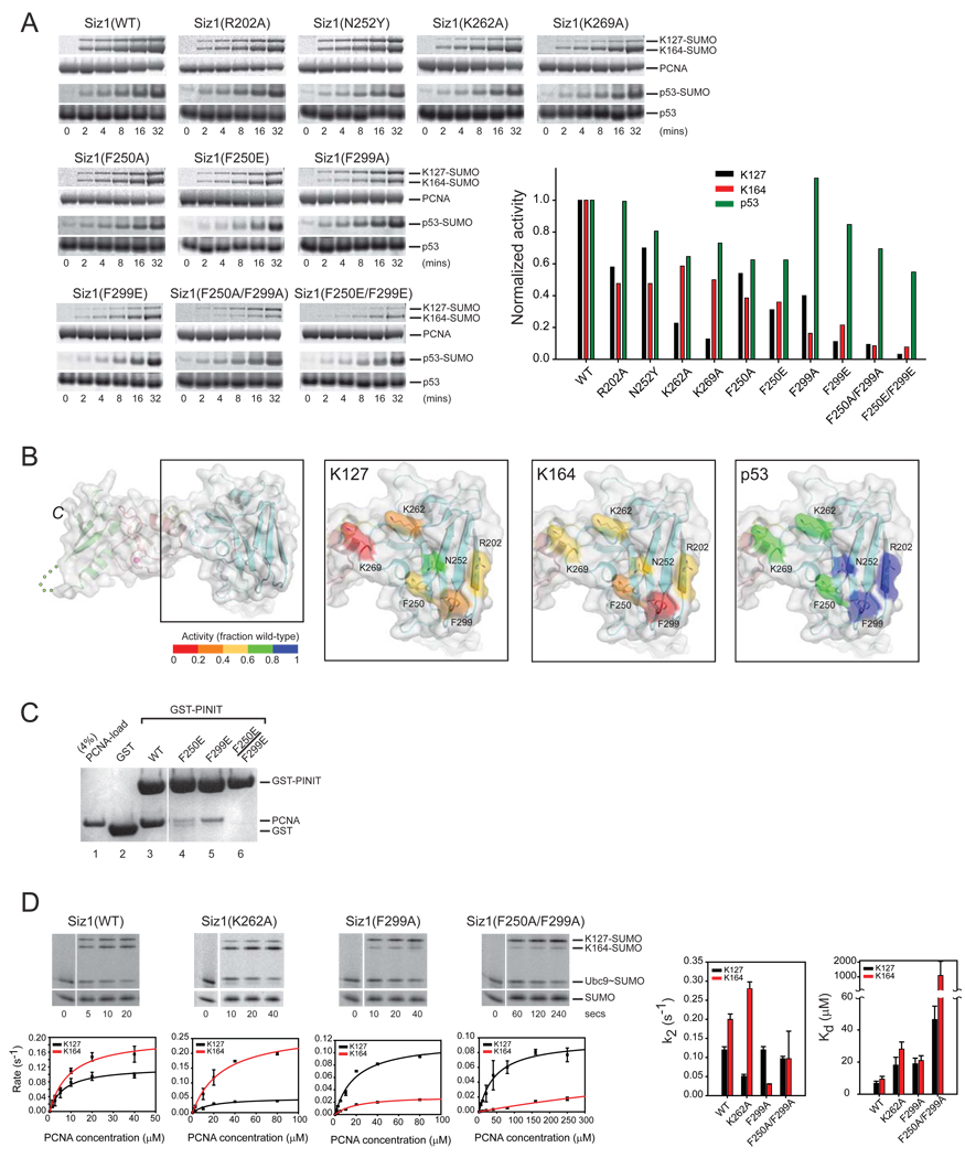
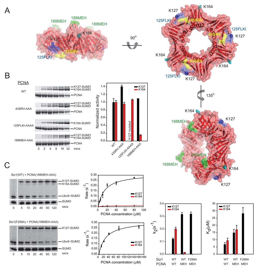
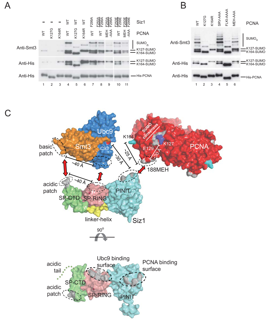
Siz1 is a founding member of the Siz/PIAS RING family of SUMO E3 ligases. The X-ray structure of an active Siz1 ligase revealed an elongated tripartite architecture comprised of an N-terminal PINIT domain, a central zinc-containing RING-like SP-RING domain, and a C-terminal domain we term the SP-CTD. Structure-based mutational analysis and biochemical studies show that the SP-RING and SP-CTD are required for activation of the E2 approximately SUMO thioester, while the PINIT domain is essential for redirecting SUMO conjugation to the proliferating cell nuclear antigen (PCNA) at lysine 164, a nonconsensus lysine residue that is not modified by the SUMO E2 in the absence of Siz1. Mutational analysis of Siz1 and PCNA revealed surfaces on both proteins that are required for efficient SUMO modification of PCNA in vitro and in vivo.


Figures

Figure 1
Figure 1. Siz/PIAS E3 ligase family members and structure of the Siz1 catalytic domain
(A) SP-RING family E3 ligases from S. cerevisiae (Siz1, Siz2), H. sapiens (PIAS1, PIASxβ, PIAS3, and PIASy) and the related SP-RING ligase Mms21. Amino acid numbers and lines indicate boundaries between domains: SAP (dark green), PINIT (cyan), linker helix (yellow), SPRING (salmon), SP-CTD (light green), acidic domain (red) and the SIM motif (blue). (B) The Siz1 structure in ribbon representation enveloped by a transparent molecular surface (top panel) with domains colored as above. Secondary structural elements are labeled and numbered. Zn2+ is depicted as a pink sphere. Each disordered amino acid residue in the SP-CTD is indicated by a green dot. Termini are indicated as N and C, respectively. Bottom panel depicts structures of the separated Siz1 SP-CTD, SP-RING, and PINIT domains.
Figure 2
Figure 2. Siz1 E3 ligase activities and deletion of the PINIT and SIM domains
(A) Schematic of Siz1 as in Figure 1A with solid bars indicating termini for proteins used in the assays below. SDS-PAGE for SUMO conjugation to PCNA (top), p53 (middle) and RanGAP1 (bottom) using Siz1(167–465), Siz1(167–479) or Siz1(167–508). (B) Similar to (A) for SUMO conjugation assays using Siz1(315–479) or Siz1(315–508). Each lane depicts the products after 1 hour at 37°C with Siz1 at the indicated concentrations. Proteins detected by SYPRO Ruby. Substrates and products are labeled.
Figure 3
Figure 3. Sequence alignment of SP-RING and SP-CTD and a structural model for SP-RING:Ubc9
(A) Amino acid alignment for SP-RING and SP-CTD domains from indicated Siz/PIAS family members with acidic residues C-terminal to the SP-CTD colored red. Structure-based sequence alignment for SP-RING, c-Cbl RING, and CHIP U-box (below). Secondary structural elements are labeled and shown above the alignment for Siz1 (color coded as in Figure 1) or below the alignment for c-Cbl RING (magenta) and CHIP U-box (gold). Circles indicate Siz1 residues subjected to mutational analysis. Asterisks indicate residues that contact the E2s in c-Cbl RING (magenta) and CHIP U-box (gold). (B) Structures of the c-Cbl RING:UbcH7 (left; magenta; PDB code 1FBV), CHIP U-box:Ubc13 (middle; gold; PDB code 2C2V) and SP-RING (salmon) with Ubc9 modeled (right) as described in the text. UbcH7, Ubc13 and Ubc9 in stick representation (blue). Residues in the E2:E3 interface in are shown in stick representation and labeled. Zn2+ ions are shown as pink spheres.
Figure 4
Figure 4. SP-RING, SP-CTD and SUMO surfaces contribute to Siz1-dependent SUMO conjugation of PCNA
(A) SDS-PAGE and time course (left) and graph depicting rates in the linear range of the assay (right). Rates normalized to one to enable a comparative analysis for SUMO conjugation to p53, PCNA at K127 and K164 with wild-type and mutant Siz1 isoforms. Proteins detected and quantified by SYPRO Ruby (Methods). (B) SP-RING and SP-CTD domains of Siz1 with amino acids subjected to mutational analysis in stick representation and Siz1 surface color-coded according to the level of activity (inset color bar) for SUMO conjugation to PCNA Lys127 (second from left), PCNA Lys164 (second from right), and p53 (right panel). (C) SDS-PAGE and time course for SUMO conjugation to PCNA with wild-type or mutant SUMO isoforms in the absence of Siz1 with 5 µM Ubc9 (top) or presence of Siz1 with 100 nM Ubc9 (bottom). (D) SDS-PAGE and time course for SUMO conjugation to PCNA with wild-type or mutant Siz1 isoforms under conditions of single turnover (left). Proteins detected by fluorescence (Alexa Fluor 488-SUMO; Methods). The initial rate of reaction (s−1) versus PCNA concentration (µM) is shown below each gel. Bar charts (right) for Kd (µM) and k2 (s−1) for indicated Siz1 isoforms. Error bars represent ±1 standard deviation.
Figure 5
Figure 5. Surfaces on the Siz1 PINIT domain are important for SUMO conjugation to PCNA
(A) SDS-PAGE and time course (left) and graph depicting rates in the linear range of the assay (right). Rates normalized to one to enable a comparative analysis for SUMO conjugation to p53, PCNA at K127 and K164 with wild-type and mutant Siz1 isoforms. Proteins detected and quantified by SYPRO Ruby (Methods). (B) PINIT domain of Siz1 with amino acids subjected to mutational analysis in stick representation and Siz1 surface color-coded according to the level of activity (inset color bar) for SUMO conjugation to PCNA Lys127 (second from left), PCNA Lys164 (second from right), and p53 (right panel). (C) SDS-PAGE of pull-down assays to probe interactions between PCNA, GST, or GST fusions to the Siz1 PINIT or indicated mutant isoforms. Protein detected by Coomassie blue (Methods). (D) SDS-PAGE and time course (left) for SUMO conjugation to PCNA with indicated Siz1 isoforms under conditions of single turnover with 10 µM PCNA (near the apparent Kd for Lys164). Proteins detected by fluorescence (Alexa Fluor 488-SUMO; Methods). The initial rate of reaction (s−1) versus PCNA concentration (µM) is shown below each gel. Bar charts (right) for Kd (µM) and k2 (s−1) for indicated Siz1 isoforms. Error bars represent ±1 standard deviation.
Figure 6
Figure 6. A PCNA surface is important for Siz1-mediated SUMO conjugation
(A) Surface representations of PCNA depicting loops 43-SRV (yellow), 125-FLKI (blue) and 188-MEH (green). PCNA Lys127 and Lys164 colored blue and cyan, respectively. (B) SDS-PAGE and time course (left) and graph depicting rates in the linear range of the assay (right) for SUMO conjugation to PCNA Lys127 and Lys164 with wild-type Siz1. Rates normalized to one for wild-type PCNA to enable a comparative analysis to PCNA mutant isoforms. Proteins detected by SYPRO Ruby (Methods). (C) SDS-PAGE and time course (left) for SUMO conjugation to PCNA(188-MEH-AAA) with Siz1(WT) or Siz1(F299A) under conditions of single turnover with 10 µM PCNA. Proteins detected by fluorescence (Alexa Fluor 488-SUMO; Methods). The initial rate of reaction (s−1) versus PCNA concentration (µM) is shown below each gel. Bar charts (right) for Kd (µM) and k2 (s−1) for indicated PCNA and Siz1 isoforms. Error bars represent ±1 standard deviation.
Figure 7
Figure 7. Surfaces on Siz1 and PCNA important for SUMO conjugation in vitro are important in vivo and a model for E2~SUMO activation and selection of PCNA Lys164
(A) Western blot to detect His-tagged PCNA and SUMO conjugated PCNA isolated from yeast cultures treated with MMS (Methods). Siz1 and PCNA alleles are indicated above each lane. (B) Same as (A) except that the yeast strain (pol30Δ) expressed wild-type Siz1 from the genomic locus and was complemented with PCNA (WT, K127G, K164R, 43SRV-AAA, 125FLKI-AAAA or 188MEH-AAA). Anti-His antibody used to detect PCNA and SUMO conjugates (middle and bottom). An anti-Smt3 antibody was used to confirm the identity of SUMO conjugated PCNA (top). (C) Model for an activated E3:E2~SUMO:PCNA complex with the Siz1 E3 ligase (colored as in Figure 1A), Ubc9 (blue), Smt3 (orange) and PCNA (red) (top). The Ubc9 catalytic cysteine (Cys93) is colored red, Lys164 of PCNA is colored cyan and SUMO consensus motif is colored gray with Lys127 in blue and Glu129 labeled. Surfaces identified in this study as important for SUMO conjugation by mutational analysis of Siz1 and PCNA are colored gray. Red arrows and dashed ellipses indicate putative pair-wise interactions. Distances between protein surfaces are indicated. (Bottom panel) Siz1 shown in an orthogonal orientation to reveal surfaces proposed to interact with Smt3, Ubc9 and PCNA (colored gray indicated by ellipses).

References

    1. Ayyagari R, Impellizzeri KJ, Yoder BL, Gary SL, Burgers PM. A mutational analysis of the yeast proliferating cell nuclear antigen indicates distinct roles in DNA replication and DNA repair. Mol Cell Biol. 1995;15:4420–4429. - PMC - PubMed
    1. Bernier-Villamor V, Sampson DA, Matunis MJ, Lima CD. Structural basis for E2-mediated SUMO conjugation revealed by a complex between ubiquitin-conjugating enzyme Ubc9 and RanGAP1. Cell. 2002;108:345–356. - PubMed
    1. Branzei D, Vanoli F, Foiani M. SUMOylation regulates Rad18-mediated template switch. Nature. 2008;456:915–920. - PubMed
    1. Cheng CH, Lo YH, Liang SS, Ti SC, Lin FM, Yeh CH, Huang HY, Wang TF. SUMO modifications control assembly of synaptonemal complex and polycomplex in meiosis of Saccharomyces cerevisiae. Genes Dev. 2006;20:2067–2081. - PMC - PubMed
    1. DeLano WL. The PyMOL Molecular Graphics System. San Carlos, CA, USA: DeLano Scientific; 2002.

Publication types

MeSH terms

Substances

Associated data

LinkOut - more resources