. 2010 Jan 15;396(2):223-30.
doi: 10.1016/j.ab.2009.09.012. Epub 2009 Sep 11.

A nonradioactive plate-based assay for stimulators of nonspecific DNA nicking by HIV-1 integrase and other nucleases

Affiliations

A nonradioactive plate-based assay for stimulators of nonspecific DNA nicking by HIV-1 integrase and other nucleases

Malgorzata Sudol et al. Anal Biochem. .

Abstract

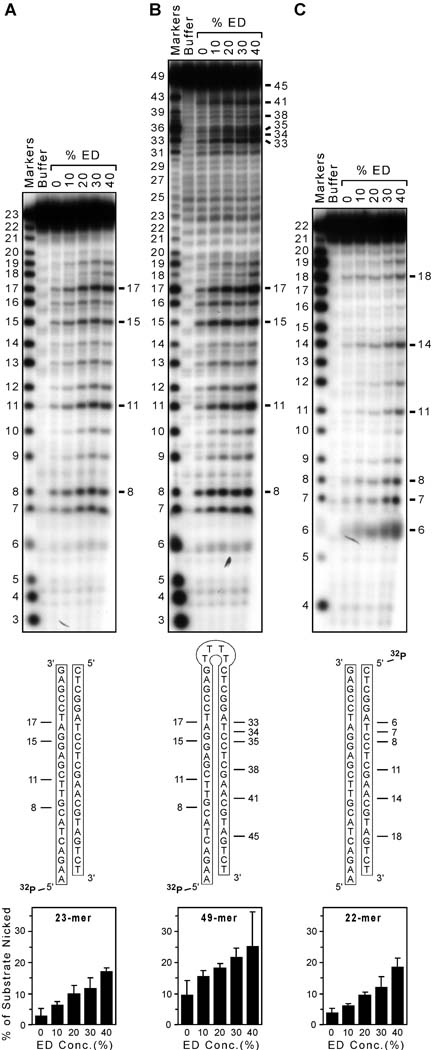
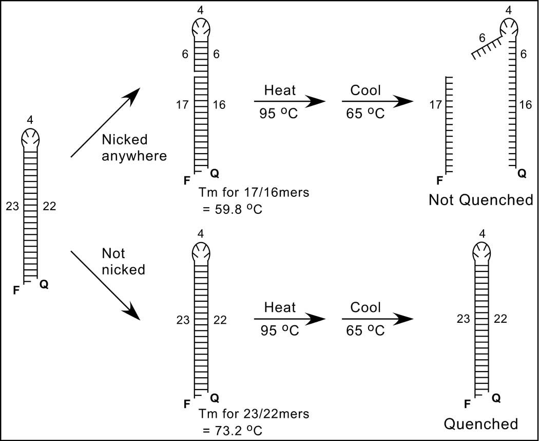
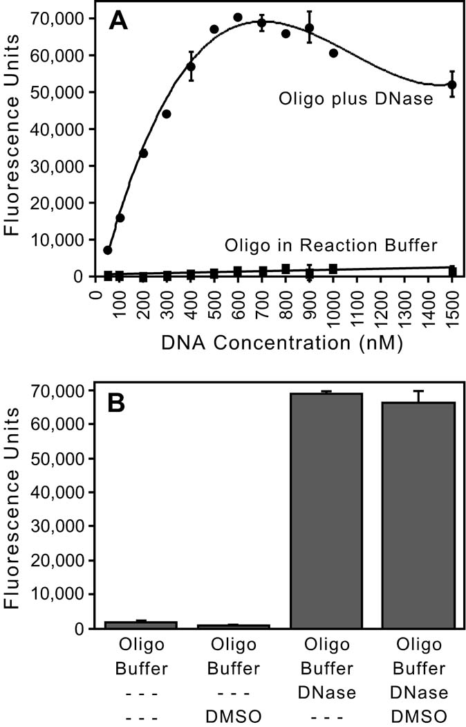
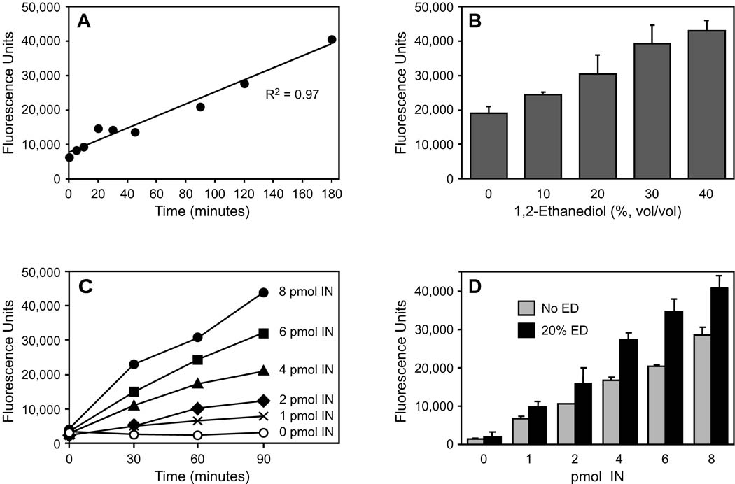
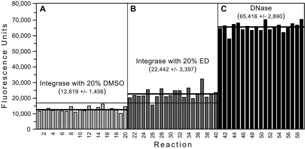
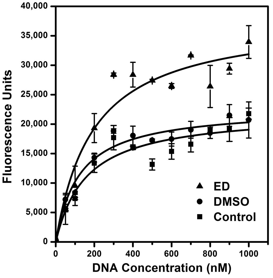
Retroviral integrase enzymes have a nonspecific endonuclease activity that is stimulated by certain compounds, suggesting that integrase could be manipulated to damage viral DNA. To identify integrase stimulator (IS) compounds as potential antiviral agents, we have developed a nonradioactive assay that is suitable for high-throughput screening. The assay uses a 49-mer oligonucleotide that is 5'-labeled with a fluorophore, 3'-tagged with a quencher, and designed to form a hairpin that mimics radioactive double-stranded substrates in gel-based nicking assays. Reactions in 384-well plates are analyzed on a real-time PCR machine after a single heat denaturation and subsequent cooling to a point between the melting temperatures of unnicked substrate and nicked products (no cycling is required). Under these conditions, unnicked DNA reforms the hairpin and quenches fluorescence, whereas completely nicked DNA yields a large signal. The assay was linear with time, stimulator concentration, and amount of integrase, and 20% concentrations of the solvent used for many chemical libraries did not interfere with the assay. The assay had an excellent Z' factor, and it reliably detected known IS compounds. This assay, which is adaptable to other nonspecific nucleases, will be useful for identifying additional IS compounds to develop the novel antiviral strategy of stimulating integrase to destroy retroviral DNA.


Figures

FIG. 1
FIG. 1. Gel-based nonspecific nicking assay
(A) Autoradiogram of nicking assays. A nonspecific 23-mer was 5' labeled and annealed to a complementary 22-mer, then incubated with protein buffer as a negative control (the second lane) or HIV-1 integrase with 0, 10, 20, 30, or 40% 1,2-ethanediol (the final 5 lanes). The buffer lane also contained 20% ED, and the first lane shows markers. Nucleotide sizes are indicated at the left, and prominent products are highlighted at the right; no products were evident near the bottom of the gel (not shown). The sequence of the 5' 32P-labeled substrate is depicted beneath the gel, and prominent nicks in the labeled strand are indicated. Two similar experiments were conducted and analyzed as in Materials and methods, and the graph at the bottom shows the percentage of substrate that was nicked as a function of the ED concentration in reactions with integrase (data are mean ± standard error). (B) Similar to panel A but using a 5' 32P-labeled 49-mer designed to form a hairpin as the substrate. (C) Similar to panel A but with the 5' 32P label on the 22-mer strand. (R2 ≥ 0.96 for the regression line for each graph at the bottom, not shown.)
FIG. 2
FIG. 2. Nonradioactive nonspecific nicking assay
A schematic of the solution-based assay is shown. The 49-mer oligonucleotide substrate at the left, which has the sequence shown in Fig. 1B and was designed to form a hairpin, is 5'-labeled with a fluorophore (F) and 3'-tagged with a quencher (Q); numbers refer to the lengths of structural features in nucleotides, and the 5' overhang permits a nontagged version to be 32P-radiolabeled for gel-based assays. The upper scheme shows that nicking on either side of the hairpin (position 17 is used as an example) will separate the F and Q groups, especially after heat-denaturation at 95°C and subsequent cooling to 65°C, which is above the melting temperature (Tm) of potential base-paired sequences (the Tm for the 17-mer and its complementary 16-mer was estimated as 59.8°C). In contrast, the lower scheme shows that unnicked DNA should reanneal and quench because the Tm of the full stem (estimated as 73.2°C) is higher than 65°C.
FIG. 3
FIG. 3. Validation of the fluorescence-based assay
(A) Effect of DNA concentration. Increasing concentrations of the F/Q-tagged oligonucleotide depicted in Fig. 2 were incubated in integrase reaction buffer or in the same buffer plus 50 µg DNase I, and reactions were conducted and analyzed as in Table 1 (except the cooling temperature in this early experiment was 63°C). The means ± SD of duplicate reactions are shown. (B) Effect of DMSO. 500 nM of the F/Q-tagged 49-mer was incubated in reaction buffer without or with 20% DMSO (the first 2 bars, respectively) or in reaction buffer plus 1 µg DNase I without or with 20% DMSO (the last 2 bars, respectively), and analyzed as in Table 1; the means ± SD of quadruplicate reactions are shown. Figs. A and B are representative of at least two experiments for each condition.
FIG. 4
FIG. 4. Parameters of the fluorescence assay
Except as noted, all reactions were conducted and analyzed as in Materials and methods. (A) Time course of integrase-mediated nicking. 500 nM of the F/Q-tagged 49-mer was incubated with 4 pmol HIV-1 integrase and 20% ED in replicate microcentrifuge tubes, and at various times individual reactions were heated at 65°C to inactivate integrase and then transferred to a 384-well plate for analysis. (B) Dose-dependant stimulation by 1,2-ethanediol. 500 nM of the F/Q-tagged 49-mer was incubated for 90 minutes with 4 pmol HIV-1 integrase and the indicated concentrations of ED; the means ± SD of triplicate reactions are shown (R2 = 0.99 for the regression line, not shown). (C) Effect of integrase concentration. 500 nM of the F/Q-tagged 49-mer was incubated with increasing amounts of HIV-1 integrase in the presence of 20% ED in microcentrifuge tubes, and at various times aliquots were removed, heated at 65°C, then transferred to a 384-well plate for analysis. (D) Stimulation of varying integrase concentrations by ED. 500 nM of the F/Q-tagged 49-mer was incubated for 90 minutes with increasing amounts of HIV-1 integrase with or without 20% ED present; the means ± SD of triplicate reactions are shown (R2 = 0.98 for each regression line, not shown; the p-values comparing the bars were 0.04 for 1 pmol integrase, 0.08 for 2 pmol integrase, < 0.002 for 4 or 6 pmol integrase, and < 0.006 for 8 pmol integrase). The number of pmols indicated in Figs. C and D is per 10-ul reaction.
FIG. 5
FIG. 5. Performance of the assay
Using the optimized conditions, 3 sets of 20 reactions were set up with: (A) 4 pmol integrase plus 20% DMSO as negative controls for baseline nicking, (B) 4 pmol integrase plus 20% ED as an example of a known integrase stimulator, and (C) 10 µg of DNase I as positive controls for maximum nicking (one reaction from the third set is not shown and was excluded from analysis because it had minimal fluorescence and apparently did not receive DNase). Reactions were conducted and analyzed as in Table 1. The mean ± SD is shown for each set of reactions, thick lines are drawn at each mean value, and the thin horizontal line indicates the mean + 3 SD for the negative controls.
FIG. 6
FIG. 6. Kinetic analysis
Reactions were set up and analyzed as in Materials and methods using optimized conditions (90 minutes and 4 pmol integrase) but the amount of substrate DNA was varied. Three reaction series (containing 20% ED, 20% DMSO, or extra water as a control) were compared. The experiment was repeated on 3 separate days, and the means ± standard errors are shown. The Vmax and Km data were calculated and the curves were fit using the OriginPro 8 program (OriginLab Corporation, Northampton, MA).

Similar articles

Cited by

References

    1. Craigie R. HIV integrase, a brief overview from chemistry to therapeutics. J. Biol. Chem. 2001;276:23213–23216. - PubMed
    1. Brown PO. Integration. In: Coffin JM, Hughes SH, Varmus HE, editors. Retroviruses. Cold Spring Harbor, New York: Cold Spring Harbor Laboratory Press; 1997. pp. 161–203.
    1. Havlir DV. HIV integrase inhibitors--out of the pipeline and into the clinic. N. Engl. J. Med. 2008;359:416–418. - PubMed
    1. Katzman M, Sudol M. Nonspecific alcoholysis, a novel endonuclease activity of human immunodeficiency virus type 1 and other retroviral integrases. J. Virol. 1996;70:2598–2604. - PMC - PubMed
    1. Skinner LM, Sudol M, Harper AL, Katzman M. Nucleophile selection for the endonuclease activities of human, ovine, and avian retroviral integrases. J. Biol. Chem. 2001;276:114–124. - PubMed

Publication types

MeSH terms

LinkOut - more resources