. 2009 Nov 27;389(4):586-92.
doi: 10.1016/j.bbrc.2009.09.025. Epub 2009 Sep 11.

The plasmodium receptor for activated C kinase protein inhibits Ca(2+) signaling in mammalian cells

Affiliations

The plasmodium receptor for activated C kinase protein inhibits Ca(2+) signaling in mammalian cells

Robson Sartorello et al. Biochem Biophys Res Commun. .

Abstract

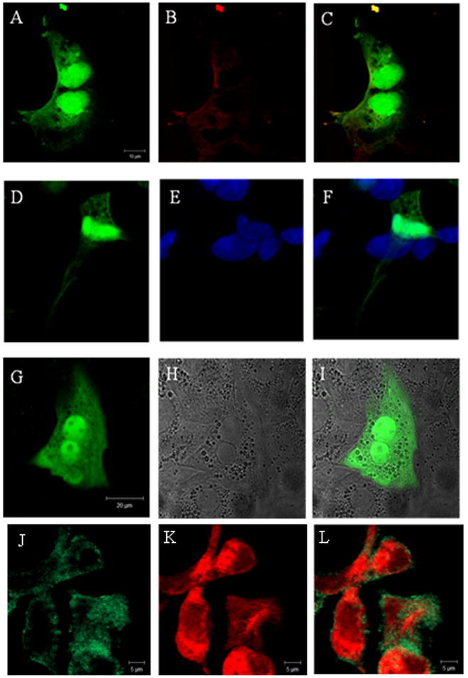
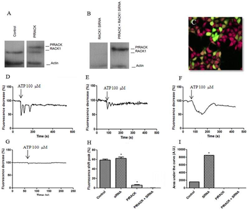
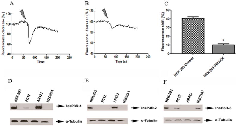
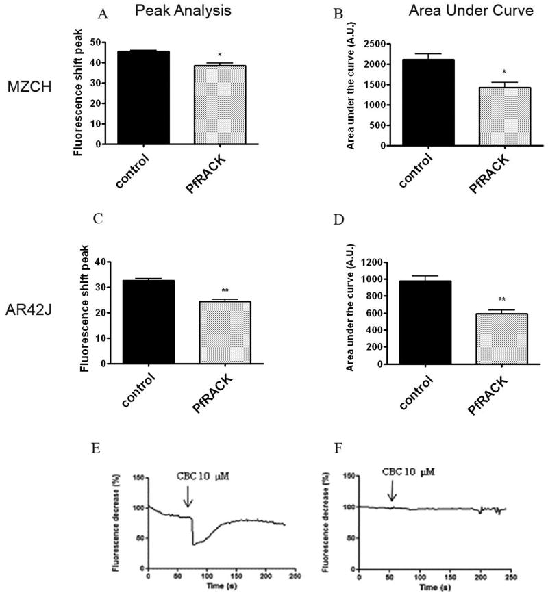
Plasmodium falciparum, the most lethal malarial parasite, expresses an ortholog for the protein kinase C (PKC) activator RACK1. However, PKC has not been identified in this parasite, and the mammalian RACK1 can interact with the inositol 1,4,5-trisphosphate receptor (InsP3R). Therefore we investigated whether the Plasmodium ortholog PfRACK also can affect InsP3R-mediated Ca(2+) signaling in mammalian cells. GFP-tagged PfRACK and endogenous RACK1 were expressed in a similar distribution within cells. PfRACK inhibited agonist-induced Ca(2+) signals in cells expressing each isoform of the InsP3R, and this effect persisted when expression of endogenous RACK1 was reduced by siRNA. PfRACK also inhibited Ca(2+) signals induced by photorelease of caged InsP3. These findings provide evidence that PfRACK directly inhibits InsP3-mediated Ca(2+) signaling in mammalian cells. Interference with host cell signaling pathways to subvert the host intracellular milieu may be an important mechanism for parasite survival.


Figures

Fig. 1
Fig. 1
Confocal immunofluorescence shows that (a) PfRACK and (b) RACK1 partially co-localizes in HEK293 cells. (c) Merged image. HEK293 cells (d) expressing GFP-PfRACK and (e) labeled with TO-PRO-3 show that (f) some PfRACK is in the nucleus. Transfection of rat hepatocytes with GFP-PfRACK shows the distribution of this protein is similar in these cells: (g) GFP fluorescence; (h) transmitted light image; (i) merged image. Mz-Cha-1 cells (j) expressing GFP-PfRACK and (k) labeled with an anti- type III InsP3R antibody show that (l) PfRACK only partially co- localizes with this InsP3R isoform.
Fig. 2
Fig. 2
Immunoblot shows (a) (from left to right) a ∼50 Kd band corresponding to RACK1 in control cells, an additional ∼75 Kd band in cells transfected with GFP-PfRACK. Immunoblot shows (b) neither band in cells treated with RACK1 siRNA, and only the ∼75 Kd band in cells treated with RACK1 siRNA and transfected with GFP-PfRACK. (c-i) Confocal image demonstrates fura-red fluorescence (red) monitored in GFP-PfRACK transfected (green) and non-transfected cells simultaneously. Typical tracings of Ca2+ signals in individual (d) non-transfected and (e) transfected cells, each stimulated with ATP (100μM). Fura-red fluorescence decreases when cytosolic Ca2+ increases. Typical tracings of Ca2+ signals in individual cells treated with siRNA to decrease expression of endogenous RACK1 reveal that (f) the ATP-induced Ca2+ signal is more sustained in cells lacking RACK1, but (g) expression of PfRACK suppresses this signal. (h) Summary of peak amplitude of ATP-induced Ca2+ signals shows that loss of RACK1 accentuates the Ca2+ signal, while Ca2+ is markedly decreased by PfRACK expression (*p<0.001). Values are mean±SEM of triplicate measurements made of >40 cells under each condition. (i) Summary of the area under the curve of ATP-induced Ca2+ signals shows that loss of RACK1 accentuates this measure of the Ca2+ signal, while it is markedly decreased by expression of PfRACK. Values are mean±SEM of triplicate measurements made of >40 cells under each condition (*p<0.001). “Control” represents cells transfected with scrambled sequence.
Fig. 3
Fig. 3
(a-c) Cells were loaded with fura-red and caged InsP3, then monitored by confocal microscopy as InsP3 was uncaged by two-photon flash-photolysis. Typical tracings of the InsP3-induced Ca2+ signal in (a) a non-transfected control cell and (b) a cell expressing GFP-PfRACK. (c) Summary shows the peak amplitude of the InsP3-induced Ca2+ signal is reduced (*p<0.001) in cells expressing GFP-PfRACK. Values are mean±SEM of triplicate measurements made of >40 cells under each condition. (d-f) Expression of InsP3R isoforms in different cell lines. Blots for (d) type I, (e) type II, and (f) type III InsP3R show that PC-12 cells express types I and III but not type II InsP3R, AR4-2J cells express types I and II but not type III InsP3R, and Mz-Cha-1 cells express only type III InsP3R. HEK293 cells express all three isoforms.
Fig. 4
Fig. 4
Ca2+ signaling was measured in cells loaded with fura-red and then examined by confocal microscopy. (a-b) Both the amplitude and area under the curve of carbachol (10μM)-induced Ca2+ signals was significantly reduced in Mz-Cha-1 cells transfected with GFP-PfRACK. (c-d) Both the amplitude and area under the curve of carbachol (10μM)-induced Ca2+ signals was significantly reduced in AR4-2J cells transfected with GFP-PfRACK. Values obtained in Mz-Cha-1 and AR4-2J cells are mean±SEM of triplicate measurements made of >40 cells under each experimental condition (*p<0.001). Typical tracings of Ca2+ signals in individual (e) non-transfected and (f) transfected PC-12 cells, each stimulated with carbachol (10μM) shows that PfRACK inhibits Ca2+ signals in these cells as well. Tracings are representative of those observed in >20 cells under each condition.

References

    1. Billker O, Dechamps S, Tewari R, Wenig G, Franke-Fayard B, Brinkmann V. Calcium and a calcium-dependent protein kinase regulate gamete formation and mosquito transmission in a malaria parasite. Cell. 2004;117:503–514. - PubMed
    1. Green JL, Rees-Channer RR, Howell SA, Martin SR, Knuepfer E, Taylor HM, Grainger M, Holder AA. The Motor Complex of Plasmodium falciparum Phosphorylation by A Calcium-Dependent Protein Kinase. Journal of Biological Chemistry. 2008;283:30980–30989. - PMC - PubMed
    1. Vaid A, Sharma P. PfPKB, a protein kinase B-like enzyme from Plasmodium falciparum II. Identification of calcium/calmodulin as its upstream activator and dissection of a novel signaling pathway. Journal of Biological Chemistry. 2006;281:27126–27133. - PubMed
    1. Vaid A, Sharma P. PfPKB, a protein kinase B-like enzyme from Plasmodium falciparum II. Identification of calcium/calmodulin as its upstream activator and dissection of a novel signaling pathway. Journal of Biological Chemistry. 2006;281:27126–27133. - PubMed
    1. Hotta CT, Gazarini ML, Beraldo FH, Varotti FP, Lopes C, Markus RP, Pozzan T, Garcia CRS. Calcium-dependent modulation by melatonin of the circadian rhythm in malarial parasites. Nature Cell Biology. 2000;2:466–468. - PubMed

Publication types

MeSH terms

Substances