. 2009 Oct 30;284(44):30148-58.
doi: 10.1074/jbc.M109.002832. Epub 2009 Sep 10.

Crowded cell-like environment accelerates the nucleation step of amyloidogenic protein misfolding

Affiliations

Crowded cell-like environment accelerates the nucleation step of amyloidogenic protein misfolding

Zheng Zhou et al. J Biol Chem. .

Abstract

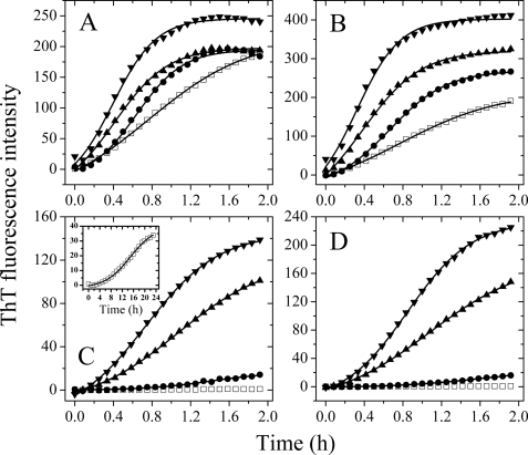
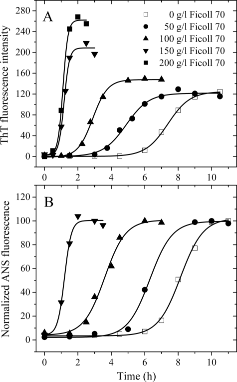
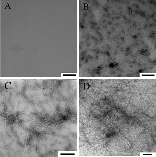
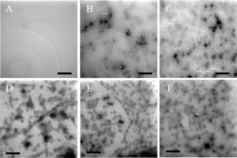
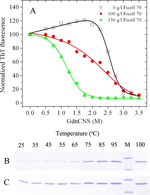
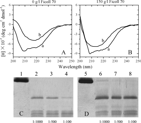
To understand the role of a crowded physiological environment in the pathogenesis of neurodegenerative diseases, we report the following. 1) The formation of fibrous aggregates of the human Tau fragment Tau-(244-441), when hyperphosphorylated by glycogen synthase kinase-3beta, is dramatically facilitated by the addition of crowding agents. 2) Fibril formation of nonphosphorylated Tau-(244-441) is only promoted moderately by macromolecular crowding. 3) Macromolecular crowding dramatically accelerates amyloid formation by human prion protein. A sigmoidal equation has been used to fit these kinetic data, including published data of human alpha-synuclein, yielding lag times and apparent rate constants for the growth of fibrils for these amyloidogenic proteins. These biochemical data indicate that crowded cell-like environments significantly accelerate the nucleation step of fibril formation of human Tau fragment/human prion protein/human alpha-synuclein (a significant decrease in the lag time). These results can in principle be predicted based on some known data concerning protein concentration effects on fibril formation both in vitro and in vivo. Furthermore, macromolecular crowding causes human prion protein to form short fibrils and nonfibrillar particles with lower conformational stability and higher protease resistance activity, compared with those formed in dilute solutions. Our data demonstrate that a crowded physiological environment could play an important role in the pathogenesis of neurodegenerative diseases by accelerating amyloidogenic protein misfolding and inducing human prion fibril fragmentation, which is considered to be an essential step in prion replication.


Figures

FIGURE 1.
FIGURE 1.
Effects of macromolecular crowding on filament formation of Tau-(244–441). Filament formation of nonphosphorylated Tau-(244–441) induced by heparin in the absence and presence of Ficoll 70 (A) and dextran 70 (B), respectively, was monitored by ThT fluorescence. The crowding agent concentrations were 0 (open square), 50 g/liter (solid circle), 100 g/liter (solid triangle), and 200 g/liter (inverted solid triangle). Filament formation of GSK-3β hyperphosphorylated Tau-(244–441) in the absence and presence of Ficoll 70 (C) and dextran 70 (D), respectively, was monitored by ThT fluorescence. The long time incubation of GSK-3β-hyperphosphorylated Tau-(244–441) is shown in the inset of C. The data were fitted to a sigmoidal equation, and the solid lines represent the best fit. The corresponding parameters are summarized in supplemental Table S1.
FIGURE 2.
FIGURE 2.
Effects of macromolecular crowding on amyloid formation of human prion protein. Human prion protein in the absence and presence of Ficoll 70 was monitored by ThT fluorescence (A) and ANS fluorescence (B). The concentration of PrP was 0.50 mg/ml. The crowding agent concentrations were 0 (open square), 50 g/liter (solid circle), 100 g/liter (solid triangle), 150 g/liter (inverted solid triangle), and 200 g/liter (solid square). The data were fitted to a sigmoidal equation, and the solid lines represent the best fit. The corresponding parameters from A are summarized in supplemental Table S2.
FIGURE 3.
FIGURE 3.
Transmission electron micrographs of human Tau fragment samples at physiological pH after incubation under different conditions. Hyperphosphorylated Tau-(244–441) (A, 2-h incubation) and nonphosphorylated Tau-(244–441) (C, 2-h incubation) were incubated in the absence of a crowding agent. Hyperphosphorylated Tau-(244–441) was incubated in the presence of 300 g/liter Ficoll 70 (B, 2-h incubation), and nonphosphorylated Tau-(244–441) was incubated in the presence of Ficoll 70 at 150 g/liter (D, 2-h incubation). A 2% (w/v) uranyl acetate solution was used to negatively stain the fibrils. The scale bars represent 200 nm for A and B and 100 nm for C and D, respectively.
FIGURE 4.
FIGURE 4.
Transmission electron micrographs of human prion protein samples at physiological pH after incubation under different conditions. Prion protein samples were incubated in the absence of a crowding agent for 6 (A), 8 (B), and 12 h (C) or in the presence of 150 g/liter Ficoll 70 for 1.5 (D), 3 (E), and 6 h (F). A 2% (w/v) uranyl acetate solution was used to negatively stain the fibrils. The scale bars represent 300 nm.
FIGURE 5.
FIGURE 5.
Global denaturation of human PrP fibrils analyzed by ThT fluorescence and SDS-PAGE. Guanidine thiocyanate-induced denaturation profiles were monitored for human PrP fibrils in the absence of a crowding agent (open squares) and in the presence of 100 g/liter Ficoll 70 (solid circles) or 150 g/liter Ficoll 70 (solid triangles) (A). Amyloid fibrils, produced from human PrP incubated in the absence and presence of crowding agents for 12 h, were incubated for 1 h at 25 °C in the presence of different concentrations of GdnSCN. The concentration of GdnSCN was then adjusted to 0.35 m, followed by a ThT binding assay. The slight increase in ThT fluorescence observed in the absence of Ficoll 70 at low concentrations of denaturant was presumably due to GdnSCN-induced dissociation of co-aggregated amyloid fibrils. Stability of human PrP fibrils in the absence of a crowding agent (B) and in the presence of 150 g/liter Ficoll 70 (C) under different temperatures was determined by SDS-PAGE. Amyloid fibrils, produced from human PrP incubated in the absence and presence of crowding agents for 12 h, were incubated with 2% SDS for 5 min under each temperature used. Lane M, protein molecular weight marker, restriction endonuclease Bsp98I (25.0 kDa) and β-lactoglobulin (18.4 kDa). Proteins in the gel were visualized by Coomassie Blue staining. The band intensities reflected susceptibility of aggregates to thermal solubilization.
FIGURE 6.
FIGURE 6.
Secondary structural changes of human PrP isoforms monitored by far-UV CD (A and B) and proteinase K digestion assays of human PrP fibrils (C and D) formed in the absence of a crowding agent (A and C) and in the presence of 150 g/liter Ficoll 70 (B and D). Curve a, native human PrP. Curve b, human PrP fibrils. Samples were treated with PK for 1 h at 37 °C at PK:PrP molar ratios as follows: 1:1000 (lanes 2 and 6), 1:500 (lanes 3 and 7), and 1:100 (lanes 4 and 8). The controls with zero protease in the absence of a crowding agent and in the presence of 150 g/liter Ficoll 70 were loaded on lanes 1 and 5, respectively. Amyloid fibrils were produced from human PrP incubated in the absence and presence of crowding agents for 12 h. Protein fragments were separated by SDS-PAGE and detected by silver staining.

References

    1. Dobson C. M. (1999) Trends Biochem. Sci. 24, 329–332 - PubMed
    1. Dobson C. M. (2003) Nature 426, 884–890 - PubMed
    1. Goedert M. (1993) Trends Neurosci. 16, 460–465 - PubMed
    1. Prusiner S. B. (1998) Proc. Natl. Acad. Sci. U.S.A. 95, 13363–13383 - PMC - PubMed
    1. Soto C. (2003) Nat. Rev. Neurosci. 4, 49–60 - PubMed

Publication types