. 2009 Oct 1;183(7):4545-53.
doi: 10.4049/jimmunol.0900673. Epub 2009 Sep 11.

Temporal regulation of Ig gene diversification revealed by single-cell imaging

Affiliations

Temporal regulation of Ig gene diversification revealed by single-cell imaging

Ellen C Ordinario et al. J Immunol. .

Abstract

Rearranged Ig V regions undergo activation-induced cytidine deaminase (AID)-initiated diversification in sequence to produce either nontemplated or templated mutations, in the related pathways of somatic hypermutation and gene conversion. In chicken DT40 B cells, gene conversion normally predominates, producing mutations templated by adjacent pseudo-V regions, but impairment of gene conversion switches mutagenesis to a nontemplated pathway. We recently showed that the activator, E2A, functions in cis to promote diversification, and that G(1) phase of cell cycle is the critical window for E2A action. By single-cell imaging of stable AID-yellow fluorescent protein transfectants, we now demonstrate that AID-yellow fluorescent protein can stably localize to the nucleus in G(1) phase, but undergoes ubiquitin-dependent proteolysis later in cell cycle. By imaging of DT40 polymerized lactose operator-lambda(R) cells, in which polymerized lactose operator tags the rearranged lambda(R) gene, we show that both the repair polymerase Poleta and the multifunctional factor MRE11/RAD50/NBS1 localize to lambda(R), and that lambda(R)/Poleta colocalizations occur predominately in G(1) phase, when they reflect repair of AID-initiated damage. We find no evidence of induction of gamma-H2AX, the phosphorylated variant histone that is a marker of double-strand breaks, and Ig gene conversion may therefore proceed by a pathway involving templated repair at DNA nicks rather than double-strand breaks. These results lead to a model in which Ig gene conversion initiates and is completed or nearly completed in G(1) phase. AID deaminates ssDNA, and restriction of mutagenesis to G(1) phase would contribute to protecting the genome from off-target attack by AID when DNA replication occurs in S phase.


Figures

Figure 1
Figure 1
Regulated nuclear localization of AID. (A) AID-YFP predominantly localizes to the cytoplasm. Left, Representative images of DT40 AID-YFP stable transfectants. Nuclear pore complex (NPC) stained with antibodies (red, center), and merged DAPI image (right). Bar, 5 μm. Right, Subcellular distribution of AID-YFP analyzed with a line profile tool of the softWoRx imaging software. (B) AIDΔC-YFP predominantly localizes to the nucleus. Notations as in panel A. (C) Phosphorylation may regulate AID-YFP localization. Representative merged DAPI images of DT40 AIDT27A-YFP, AIDS38A-YFP and AIDY184A-YFP stable transfectants. (D) AID-YFP is present in the nucleus in G1 phase. Left, Representative images of DT40 AID-YFP cells treated with LMB (50 ng/ml, 1 hr), then labeled with BrdU (10 μM, 30 min). Bar, 5 μm. Right, Cell cycle distribution of cells containing nuclear AID-YFP (21%; n = 123 cells), as determined by BrdU staining and nuclear size. (E) AID-YFP may be degraded in the nucleus in S phase. Left, Representative images of DT40 AID-YFP cells treated with MG132 (50 μM, 4 hr) and LMB (50 ng/ml, 1 hr); Golgi apparatus stained with antibodies (red). Bar, 5 μm. Right, Cell cycle distribution of DT40 AID-YFP cells treated with MG132 and LMB, which contain nuclear AID-YFP (18%; n = 298 cells). (F) AID-YFP may form aggresomes following MG132 treatment. Representative images of DT40 AID-YFP cells treated with MG132 (50 μM, 4 hr) and LMB (50 ng/ml, 1 hr), and stained with antibodies to ubiquitin or SUMO (red, center); and merged DAPI image (right). Arrows indicate potential colocalization of AID-YFP and ubiquitin staining signal.
Figure 2
Figure 2
E2A preferentially colocalizes with the rearranged λR allele. (A) Schematic of the PolyLacO-tagged unrearranged Igλ locus in DT40 PolyLacO-λU cells. The ψVλ array, variable (Vλ), joining (Jλ), and constant (Cλ) regions are shown. PolyLacO is integrated between ψV17 and ψV20. (B) Proliferation of DT40, DT40 PolyLacO-λR and DT40 PolyLacO-λU cells. Cell counting and culture passage were carried out daily and growth rate was calculated as a cumulative cell number. Data shown are the average from two separate experiments. (C) Cell cycle profile of DT40, DT40 PolyLacO-λR and DT40 PolyLacO-λU cells. Exponentially growing cells were analyzed by flow cytometry after staining with propidium iodide. (D) Accumulation of sIgM-loss variants by DT40, DT40 PolyLacO-λR and DT40 PolyLacO-λU cells. Frequencies of sIgM-loss variants in 24 subclones from each line were quantitated by flow cytometry following 6 weeks of clonal expansion. Mean sIgM-loss frequencies were 0.8%, 0.9% and 0.6%, respectively, as indicated at the bottom of the panel. (E) Comparison of levels of colocalizations of E2A with the rearranged λR allele in DT40 PolyLacO-λR cells or the unrearranged λU allele in DT40 PolyLacO-λU cells.
Figure 3
Figure 3
Colocalizations of rearranged λR genes with Polη and MRN. (A) Schematic of the PolyLacO-tagged rearranged Igλ locus in DT40 PolyLacO-λR cells. The upstream ψVλ array, variable (Vλ), joining (Jλ), and constant (Cλ) regions are shown. PolyLacO is integrated between ψV17 and ψV20. (B) Left, Representative image of λR/Polη-GFP colocalization in DT40 PolyLacO-λR RFP-LacI Polη-GFP cells. Nuclear perimeter as determined by DAPI staining is outlined by the dashed white line; arrows point to colocalizations. Colocalizations were observed in 11% of cells (n = 1013), in two experiments. Right, Cell cycle distribution of total λR/Polη-GFP colocalizations; cell cycle was determined by analysis of nuclear radius (n = 79). (C) Left, Representative image of λR/NBS1 colocalization in DT40 PolyLacO-λR GFP-LacI cells. Notations as in B. Colocalizations were observed in 17% of cells (n = 232 cells), in two experiments). Right, Cell cycle distribution of total λR/NBS1 colocalizations; cell cycle was determined by analysis of nuclear radius (n = 75). (D) Above left, Representative immunofluorescent images of DT40, unirradiated (0 Gy), stained with anti-γ-H2AX (left) and anti-NBS1 (center) antibodies; merged DAPI image (right); bar, 10 μm. Above right, Percent of cells containing γ-H2AX or NBS1 foci in each stage of cell cycle; no colocalization; n = 410 cells; three experiments. Below, Representative immunofluorescent images of DT40, irradiated (8 Gy, 2 hrs post IR) and stained as above; 38% colocalization; n = 312 cells; three experiments.
Figure 4
Figure 4
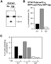
UNG-dependent localizations of Polη-GFP at rearranged λR genes. (A) Uracil-DNA excision activity in the nuclear extract of DT40 PolyLacO-λR RFP-LacI Polη-GFP cells or its derivative stably expressing Ugi (Ugi − and +, respectively). (B) Cell cycle distribution of λR/Polη-GFP colocalizations in DT40 PolyLacO-λR RFP-LacI Polη-GFP Ugi cells (n = 16); 298 cells analyzed. (C) Fraction of λR/Polη-GFP colocalizations in each stage of cell cycle, presented as the percent of total cells in which colocalizations were evident. Ugi expression diminished total colocalizations by about one-half.
Figure 5
Figure 5
Model for temporal regulation of Ig V region diversification. Diversification is activated by E2A, initiated by DNA deamination by AID and deglycosylation by UNG, generating an AP site (diamond) which is cleaved by MRN, creating a nick. The nick primes nontemplated (somatic hypermutation, left), or templated (gene conversion, right) repair DNA synthesis by Polη. Ig V region diversification may be initiated and nearly completed in G1 phase of cell cycle, producing a heteroduplex. Replication in S phase fixes mutations. If initial mutagenesis affects only a single DNA strand, then one mutated and one germline chromatid will segregate in G2 phase.

Similar articles

Cited by

References

    1. Li Z, Woo CJ, Iglesias-Ussel MD, Ronai D, Scharff MD. The generation of antibody diversity through somatic hypermutation and class switch recombination. Genes Dev. 2004;18:1–11. - PubMed
    1. Maizels N. Immunoglobulin gene diversification. Annu Rev Genet. 2005;39:23–46. - PubMed
    1. Longerich S, Basu U, Alt F, Storb U. AID in somatic hypermutation and class switch recombination. Curr Opin Immunol. 2006;18:164–174. - PubMed
    1. Martomo SA, Gearhart PJ. Somatic hypermutation: subverted DNA repair. Curr Opin Immunol. 2006;18:243–248. - PubMed
    1. Di Noia JM, Neuberger MS. Molecular mechanisms of antibody somatic hypermutation. Annu Rev Biochem. 2007;76:1–22. - PubMed

Publication types

MeSH terms

Substances