. 2009 Oct 1;183(7):4458-66.
doi: 10.4049/jimmunol.0901465. Epub 2009 Sep 11.

Engagement of TLR2 does not reverse the suppressor function of mouse regulatory T cells, but promotes their survival

Affiliations

Engagement of TLR2 does not reverse the suppressor function of mouse regulatory T cells, but promotes their survival

Qian Chen et al. J Immunol. .

Abstract

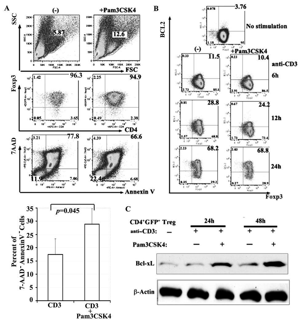
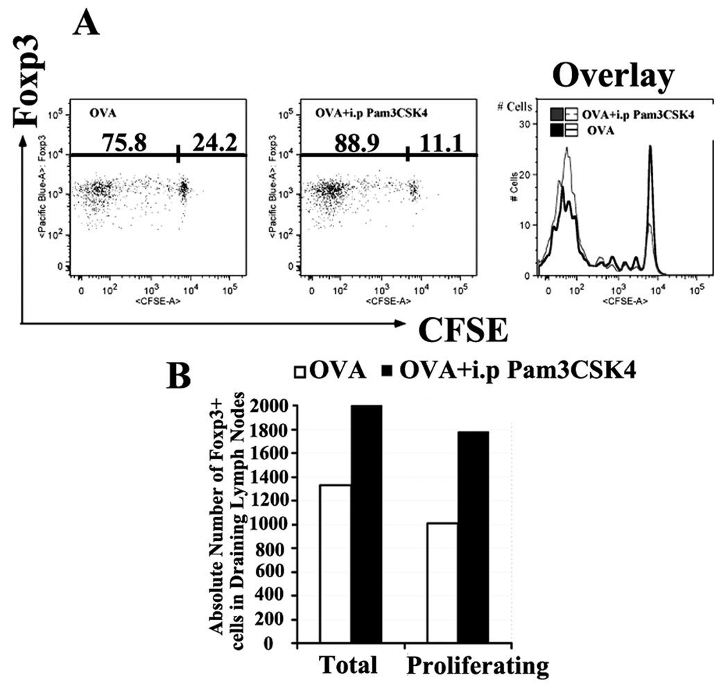
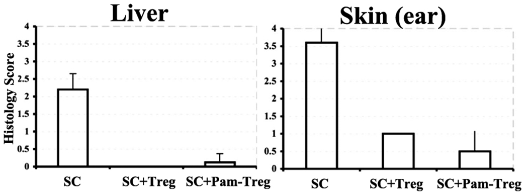
TLRs are a class of conserved pattern recognition receptors that are used by cells of the innate immune system. Recent studies have demonstrated the expression of TLRs on both human and mouse T cells raising the possibility that TLRs play a direct role in adaptive immunity. TLR2 is activated primarily by bacterial wall components including peptidoglycan and lipoproteins. Several studies have shown that mouse regulatory T (Treg) cells express TLR2 and claimed that engagement of TLR2 by synthetic ligands reversed their suppressive function. In contrary, enhancement of Treg function was observed following engagement of TLR2 on human Treg. We have reexamined the expression and function of TLR2 on mouse Treg purified from Foxp3-GFP knock-in mice. TLR2 ligation by TLR2 agonist, the synthetic bacterial lipoprotein Pam3CSK4, enhanced the proliferative responses of both conventional T cells and Treg in response to TLR stimulation in the absence of APC. Treatment of Foxp3+ Treg with Pam3CSK4 did not alter their suppressive function in vitro or in vivo and did not reduce their level of Foxp3 expression. An additional effect of TLR2 stimulation of Treg was induction of Bcl-x(L) resulting in enhanced survival in vitro. Treatment of mice with the TLR2 agonist enhanced the Ag-driven proliferation of Treg in vivo, but did not abolish their ability to suppress the development of experimental autoimmune encephalomyelitis. Development of methods to selectively stimulate TLR2 on Treg may lead to a novel approaches for the treatment of autoimmune diseases.


Conflict of interest statement

Disclosures

The authors have no financial conflict of interest.

Figures

FIGURE 1
FIGURE 1
Expression of TLR2 mRNA in GFP+ Treg and GFP T cells. A, FACS-sorted GFP T cells or GFP+ Treg (5×10⁴) were either un-stimulated or activated by plate-bound anti-CD3 in the absence or presence of Pam3CSK4 for 16h. IL-2 was added to Treg cultures. Total cellular RNA was isolated and TLR2 mRNA expression was measured by real-time PCR and normalized to β-actin mRNA levels. Epithelial cells from TLR2−/− mice served as negative control. Macrophages derived from WT mouse bone marrow following in culture in MCSF and IL-3 served as a positive control population. The TLR2 mRNA level in TLR2−/− epithelial cells was set at 1.0, and all other normalized mRNA levels were plotted relative to that value. B, The TLR2 mRNA level in un-stimulated GFP T cells was set at 1.0, and all other normalized mRNA levels were plotted relative to that value. Each point represents the mean +/− SEM of triplicate samples. Similar results were seen in one other experiment.
FIGURE 2
FIGURE 2
Effect of TLR agonists on the proliferation of GFP+ Treg and GFP T cells in vitro. A and B, FACS-sorted GFP+ Treg (5×10⁴) were cultured for 3 days with T-depleted spleen cells and soluble anti-CD3 (1µg/ml) (A) or with plate-bound anti-CD3 (5µg/ml) (B) in the presence of the following TLR agonists: Poly I:C (TLR3), CPG (TLR9), TLR7/8 (CL097) and Pam3CSK4 (TLR2-TLR1). 3H-TdR incorporation was determined during the last 6h of culture. Results are expressed as the mean +/− SEM of triplicate cultures. C and D, Same protocol as in A except IL-2 was added to all cultures. E, GFP+ Treg or F, GFP T cells were stimulated with different concentration of plate-bound anti-CD3 in the absence and presence of Pam3CSK4 (1µg/ml). Statistical analyses were performed with the nonparametric Mann-Whitney u test for the triplicate (*p<0.03; **p<0.0001). Similar results were seen in three other experiments.
FIGURE 3
FIGURE 3
Effect of different TLR2 complex agonists on the proliferation of GFP+ Treg. FACS-sorted GFP+ Treg (5×10⁴) were cultured with plate-bound anti-CD3 (5µg/ml) and IL-2 for 3 days in the presence or absence of: A, TLR2/TLR2 agonist, LTA-SA; B, TLR2/TLR6 agonist, Pam2CSK4 and C, TLR2/TLR1 agonist, Pam3CSK4. Proliferation was measured as in Figure 1. All experiments were performed at least 3 times.
FIGURE 4
FIGURE 4
Pam3CSK4 stimulation does not alter Foxp3 expression or Treg suppressive activity. A, FACS-sorted GFP+ Treg were stimulated with plate-bound anti-CD3 and IL-2 for 3 days in the presence or absence of Pam3CSK4. Flow cytometric analysis of GFP expression (right panel) and Foxp3 (left panel) was performed after the cells were rested in the presence of IL-2 alone for 2 days. B, CD4+GFP responder T cells and T-depleted spleen cells APCs (5×10⁴ of each) from TLR2−/− mice were co-cultured with the indicated number of WT GFP+ Treg and soluble anti-CD3 in the absence or presence of Pam3CSK4. T cell proliferation was assayed as in Figure 1. C, FACS-sorted GFP+ Treg from WT mice were stimulated with plate-bound anti-CD3 and IL-2 for 3 days in the presence or absence of Pam3CSK4. The indicated number of pre-activated Treg was tested for suppressive activity as in Panel B in the presence or absence of Pam3CSK4. Experiments were repeated at least 3 times.
FIGURE 5
FIGURE 5
Pam3CSK4 augments Treg survival and induces Bcl-xL but not Bcl-2 expression. A, FACS-sorted GFP+ Treg were cultured with plate-bound anti-CD3 (0.4µg/ml) and IL-2 for 7 days in the absence (left panel) or presence (right panel) of Pam3CSK4. The cells were then analyzed by flow cytometry. FSC/SSC plots and the percentage of live cells (high FSC) is indicated (top panel). Gated live cells were analyzed for GFP expression (middle panel). Viability of the total cell population was measured as the percentage of 7-AAD and Annexin V excluding cells (bottom panel). Percent of live (7-AAD AnnexinV) Treg were calculated by combined data from 3 independent experiments (below). Values of p were calculated using the Mann-Whitney test. B, Treg were stimulated with plate-bound anti-CD3 (0.4µg/ml) and IL-2 in the absence (left panel) or presence (right panel) of Pam3CSK4 for the indicated times. Bcl-2 and GFP expression were determined by flow cytometry. C, Treg were stimulated as in panel B for the indicated times. The cells were then lysed and analyzed by Western blot for mouse Bcl-xL (top panel). β-actin (bottom panel) was used as the loading control. All data were representative of three different experiments with similar results.
FIGURE 6
FIGURE 6
Pam3CSK4 enhances antigen-specific Treg proliferation in vivo. CD4+CD25+ (1×10⁶) were purified by cell sorting from OT-II-transgenic mice, CFSE-labeled and injected into TLR2−/− mice via the tail vein. 24h later, the recipient mice were immunized by s.c. injection in the flank with OVA-peptide (10µg/mouse) in IFA and simultaneously injected with either PBS or Pam3CSK4 (100µg/mouse) i.p. 5 days after immunization, the draining lymph nodes were removed and analyzed by flow cytometry for CFSE content of the transferred cells by gating on Vα2+Vβ5+Foxp3+ cells. A, Each plot indicates percentages of proliferating (CFSElow) versus resting (CFSEhigh) Treg challenged with OVA or OVA and Pam3CSK4. The overlay of CFSE histograms is shown in right panel (antigen alone, black line; antigen and Pam3CSK4, grey dashed line). B, The total versus proliferating OT-II Foxp3+ cell counts in draining lymph nodes from TLR2−/− mice were calculated using CFSE profiles. Data were representative of 2 different experiments with similar results.
FIGURE 7
FIGURE 7
Pam3CSK4-pre-treated Treg prevent the activation of Scurfy effector T cells in RAG−/− mice. Peripheral lymph node and spleen cells (5×10⁶) from 7-day-old Scurfy mice (SC) were either transferred alone into male RAG−/− mice or co-transferred with Treg (1×10⁶) that had been expanded for 3 days by stimulation with plate-bound anti-CD3 and IL-2 in the presence or absence of Pam3CSK4. Twenty-eight days after transfer, the ears and livers were evaluated histologically. Values indicate average histological score of five mice ± SD.
FIGURE 8
FIGURE 8
Pam3CSK4 treatment does not attenuate Treg-mediated prevention of EAE. Three groups (five mice in each) of TLR2−/− mice were immunized with MOG peptide/CFA on day 0 and injected with pertussis toxin on day 0 and 2 for EAE induction. One group was untreated, while the other two had received GFP+ Treg (1×10⁶) i.v. from WT mice one day before EAE induction. Pam3CSK4 (20µg/mouse) i.p. was injected to one of the groups that had received Treg on day −1, 1, 3 and 5. Mice were then monitored daily for disease until day 30. EAE clinical scores for control (filled diamonds) and for Treg-treated mice without (open squares) or with (open triangles) administration of Pam3CSK4 were shown. Data were representative of 2 different experiments with similar results.

References

    1. Marshak-Rothstein A. Toll-like receptors in systemic autoimmune disease. Nat. Rev. Immunol. 2006;6:823–835. - PMC - PubMed
    1. Takeda K, Akira S. Toll receptors and pathogen resistance. Cell. Microbiol. 2003;5:143–153. - PubMed
    1. Takeda K, Kaisho T, Akira S. Toll-like receptors. Annu. Rev. Immunol. 2003;21:335–376. - PubMed
    1. Kabelitz D. Expression and function of Toll-like receptors in T lymphocytes. Curr. Opin. Immunol. 2007;19:39–45. - PubMed
    1. Liu H, Komai-Koma M, Xu D, Liew FY. Toll-like receptor 2 signaling modulates the functions of CD4+ CD25+ regulatory T cells. Proc. Natl. Acad. Sci. U. S. A. 2006;103:7048–7053. - PMC - PubMed

Publication types

MeSH terms