. 2009 Nov;191(22):6804-11.
doi: 10.1128/JB.01006-09. Epub 2009 Sep 11.

Mutualistic biofilm communities develop with Porphyromonas gingivalis and initial, early, and late colonizers of enamel

Affiliations

Mutualistic biofilm communities develop with Porphyromonas gingivalis and initial, early, and late colonizers of enamel

Saravanan Periasamy et al. J Bacteriol. 2009 Nov.

Abstract

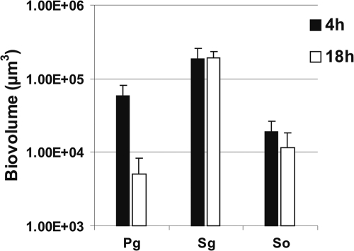
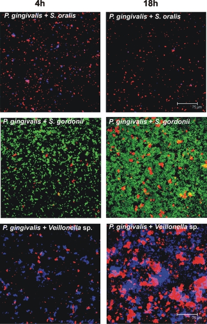
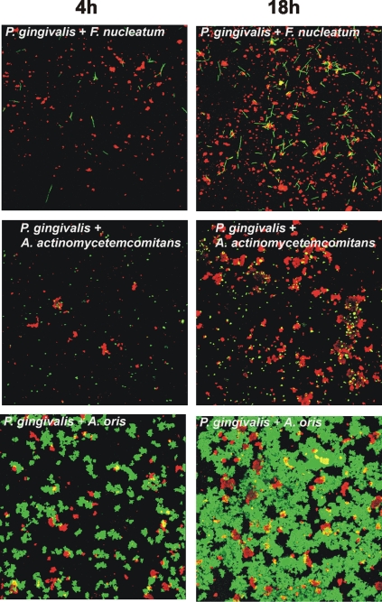
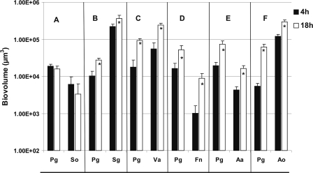
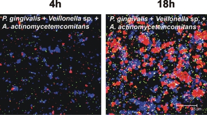
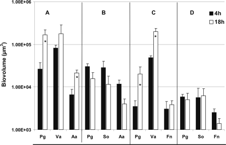
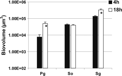
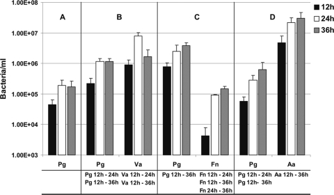
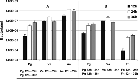
Porphyromonas gingivalis is present in dental plaque as early as 4 h after tooth cleaning, but it is also associated with periodontal disease, a late-developing event in the microbial successions that characterize daily plaque development. We report here that P. gingivalis ATCC 33277 is remarkable in its ability to interact with a variety of initial, early, middle, and late colonizers growing solely on saliva. Integration of P. gingivalis into multispecies communities was investigated by using two in vitro biofilm models. In flow cells, bacterial growth was quantified using fluorescently conjugated antibodies against each species, and static biofilm growth on saliva-submerged polystyrene pegs was analyzed by quantitative real-time PCR using species-specific primers. P. gingivalis could not grow as a single species or together with initial colonizer Streptococcus oralis but showed mutualistic growth when paired with two other initial colonizers, Streptococcus gordonii and Actinomyces oris, as well as with Veillonella sp. (early colonizer), Fusobacterium nucleatum (middle colonizer), and Aggregatibacter actinomycetemcomitans (late colonizer). In three-species flow cells, P. gingivalis grew with Veillonella sp. and A. actinomycetemcomitans but not with S. oralis and A. actinomycetemcomitans. Also, it grew with Veillonella sp. and F. nucleatum but not with S. oralis and F. nucleatum, indicating that P. gingivalis and S. oralis are not compatible. However, P. gingivalis grew in combination with S. gordonii and S. oralis, demonstrating its ability to overcome the incompatibility when cultured with a second initially colonizing species. Collectively, these data help explain the observed presence of P. gingivalis at all stages of dental plaque development.


Figures

FIG. 1.
FIG. 1.
Time-resolved change in biovolume (μm3 per field of view) of P. gingivalis (Pg), S. gordonii (Sg), and S. oralis (So) following 4 h and 18 h of incubation as single species in 25% saliva-fed flow cells.
FIG. 2.
FIG. 2.
Representative confocal micrographs of 4-h (left column) and 18-h (right column) biofilms showing no growth (top panels, P. gingivalis + S. oralis) and mutualistic growth (middle panels, P. gingivalis + S. gordonii; bottom panels, P. gingivalis + Veillonella sp.) in flow cells inoculated with two species. Bacterial cells were stained with species-specific fluorophore-conjugated immunoglobulin G (red, P. gingivalis; blue, S. oralis/Veillonella sp., green, S. gordonii), and cell-cell contact between species is evident.
FIG. 3.
FIG. 3.
Representative confocal micrographs of 4-h (left column) and 18-h (right column) biofilms showing mutualistic growth (top panels, P. gingivalis + F. nucleatum; middle panels, P. gingivalis + A. actinomycetemcomitans) and commensal growth (P. gingivalis + A. oris) in flow cells inoculated with two species. Bacterial cells were stained with species-specific fluorophore-conjugated immunoglobulin G (red, P. gingivalis; green, F. nucleatum/A. actinomycetemcomitans/A. oris), and cell-cell contact between species is evident.
FIG. 4.
FIG. 4.
Time-resolved changes in biovolumes (μm3 per field of view) of two-species flow cells inoculated with coaggregates of P. gingivalis (Pg) and S. oralis (So) (A), S. gordonii (Sg) (B), Veillonella sp. (Va) (C), F. nucleatum (Fn) (D), A. actinomycetemcomitans (Aa) (E), or A. oris (Ao) (F). An asterisk indicates statistically significant increases (P < 0.05) in bacterial growth.
FIG. 5.
FIG. 5.
Representative confocal micrographs of biofilm from three-species-inoculated flow cell. Communities following 4 h and at 18 h of incubation show intimate interaction of Veillonella sp. (blue) with A. actinomycetemcomitans (green) and P. gingivalis (red) to form mutualistic multispecies communities.
FIG. 6.
FIG. 6.
Time-resolved changes in biovolumes (μm3 per field of view) of three-species-inoculated flow cell following 4-h (black bar) and 18-h (open bar) incubation showing growth enhanced by Veillonella sp. and growth inhibited by S. oralis. (A) P. gingivalis (Pg) + Veillonella sp. (Va) + A. actinomycetemcomitans (Aa); (B) P. gingivalis (Pg) + S. oralis (So) + A. actinomycetemcomitans (Aa); (C) P. gingivalis (Pg) + Veillonella sp. (Va) + F. nucleatum (Fn); and (D) P. gingivalis (Pg) + S. oralis (So) + F. nucleatum (Fn). An asterisk indicates statistically significant increases (P < 0.05) in bacterial growth.
FIG. 7.
FIG. 7.
Time-resolved changes in biovolume (μm3 per field of view) of three-species-inoculated flow cell showing specificity by P. gingivalis (Pg) for S. gordonii (Sg) in the presence of S. oralis (So) following 4-h (black bar) and 18-h (open bar) incubation. An asterisk indicates statistically significant increases (P < 0.05) in bacterial growth.
FIG. 8.
FIG. 8.
q-PCR quantification of P. gingivalis cells alone in biofilms and with coaggregation partners in two-species-inoculated biofilms grown on polystyrene pegs submerged in 25% saliva and incubated anaerobically for 12, 24, or 36 h. (A) Single species, P. gingivalis; (B) two species, P. gingivalis (Pg) + Veillonella sp. (Va); (C) two species, P. gingivalis (Pg) + F. nucleatum (Fn); and (D) two species, P. gingivalis (Pg) + A. actinomycetemcomitans (Aa). Statistically significant increases (P < 0.05) in bacterial growth on the pegs is indicated at the bottom of each panel; for example in panel B, “Pg 12 h - 24 h” indicates that significant growth of P. gingivalis occurred between 12 h and 24 h in the two-species biofilm with Veillonella sp.
FIG. 9.
FIG. 9.
q-PCR quantification of three-species-inoculated biofilms grown on polystyrene pegs submerged in 25% saliva and incubated anaerobically for 12, 24, or 36 h. Pg, P. gingivalis; Va, Veillonella sp.; Aa, A. actinomycetemcomitans; and Fn, F. nucleatum. Statistically significant increases (P < 0.05) in bacterial growth on the pegs is indicated at the bottom of each panel; for example in panel A, “Pg 12 h - 24 h” indicates that significant growth of P. gingivalis occurred between 12 h and 24 h in the three-species inoculated biofilm with Veillonella sp. and A. actinomycetemcomitans.

References

    1. Bos, R., H. C. van der Mei, and H. J. Busscher. 1996. Co-adhesion of oral microbial pairs under flow in the presence of saliva and lactose. J. Dent. Res. 75:809-815. - PubMed
    1. Ceri, H., M. E. Olson, C. Stremick, R. R. Read, D. Morck, and A. Buret. 1999. The Calgary Biofilm Device: new technology for rapid determination of antibiotic susceptibilities of bacterial biofilms. J. Clin. Microbiol. 37:1771-1776. - PMC - PubMed
    1. Chalmers, N. I., R. J. Palmer, Jr., J. O. Cisar, and P. E. Kolenbrander. 2008. Characterization of a Streptococcus sp.-Veillonella sp. community micromanipulated from dental plaque. J. Bacteriol. 190:8145-8154. - PMC - PubMed
    1. Dawes, C., S. Watanabe, P. Biglow-Lecomte, and G. H. Dibdin. 1989. Estimation of the velocity of the salivary film at some different locations in the mouth. J. Dent. Res. 68:1479-1482. - PubMed
    1. Diaz, P. I., N. I. Chalmers, A. H. Rickard, C. Kong, C. L. Milburn, R. J. Palmer, Jr., and P. E. Kolenbrander. 2006. Molecular characterization of subject-specific oral microflora during initial colonization of enamel. Appl. Environ. Microbiol. 72:2837-2848. - PMC - PubMed

Publication types

MeSH terms

LinkOut - more resources