. 2009 Nov;75(21):6856-63.
doi: 10.1128/AEM.00540-09. Epub 2009 Sep 11.

Cryptosporidium propidium monoazide-PCR, a molecular biology-based technique for genotyping of viable Cryptosporidium oocysts

Affiliations

Cryptosporidium propidium monoazide-PCR, a molecular biology-based technique for genotyping of viable Cryptosporidium oocysts

Cristin C Brescia et al. Appl Environ Microbiol. 2009 Nov.

Abstract

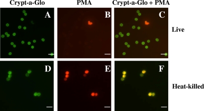
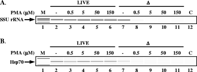
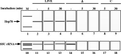
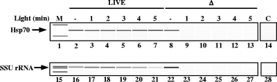
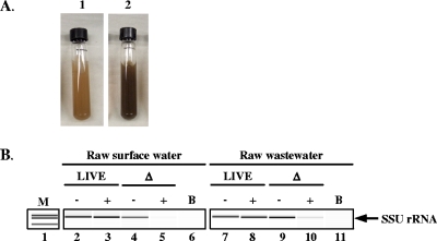
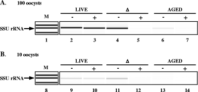
Cryptosporidium is an important waterborne protozoan parasite that can cause severe diarrhea and death in the immunocompromised. The current methods used to monitor for Cryptosporidium oocysts in water are the microscopy-based USEPA methods 1622 and 1623. These methods assess total levels of oocysts in source waters, but do not determine oocyst viability or genotype. Recently, propidium monoazide (PMA) has been used in conjunction with molecular diagnostic tools to identify species and assess the viability of bacteria. The goal of this study was the development of a Cryptosporidium PMA-PCR (CryptoPMA-PCR) assay that includes PMA treatment prior to PCR analysis in order to prevent the amplification of DNA from dead oocysts. The results demonstrated that PMA penetrates only dead oocysts and blocks amplification of their DNA. The CryptoPMA-PCR assay can also specifically detect live oocysts within a mixed population of live and dead oocysts. More importantly, live oocysts, not dead oocysts, were detected in raw waste or surface water samples spiked with Cryptosporidium oocysts. This proof-of-concept study is the first to demonstrate the use of PMA for pre-PCR treatment of Cryptosporidium oocysts. The CryptoPMA-PCR assay is an attractive approach to specifically detect and genotype viable Cryptosporidium oocysts in the water, which is critical for human health risk assessment.


Figures

FIG. 1.
FIG. 1.
Microscopic analyses distinguishing live from dead C. parvum oocysts using PMA. Live (A to C) or heat-killed (D to F) (70°C, 20 min) C. parvum oocysts treated with PMA and stained with Crypt-a-Glo. Crypt-a-Glo-labeled oocysts (green) are shown in panels A and D; the same cysts stained with PMA (red) are shown in panels B and E; and panels C and F represent overlaid images with Crypt-a-Glo and PMA (yellow) staining. Bar = 10 μm.
FIG. 2.
FIG. 2.
Effects of different PMA concentrations on detecting live and heat-killed C. parvum oocysts. Live or heat-killed C. parvum oocysts were incubated with 0.5, 5, 50, or 150 μM PMA for 10 min followed by 2 min of light exposure. This was followed by the extraction of genomic DNA and PCR amplification with primers targeting the SSU rRNA gene (A) or the hsp70 gene (B). PCR products were analyzed using an Agilent Bioanalyzer 2100. M, marker; −, no PMA control; Δ, oocysts treated at 70°C for 30 min; C, PCR control that lacks a template.
FIG. 3.
FIG. 3.
Effects of PMA incubation time on detecting live and heat-killed C. parvum oocysts. Live or heat-killed C. parvum oocysts were incubated with 150 μM PMA for either 5 or 30 min and then exposed to light for 2 min. Genomic DNA was purified as described in Materials and Methods. Amplification of a portion of the hsp70 gene (lanes 2 to 9) or the SSU rRNA gene (lanes 11 to 18) was performed using PCR. Gel electrophoresis analysis was performed using an Agilent Bioanalyzer 2100. M, marker; −, no PMA control; Δ, oocysts treated at 70°C; C, PCR control that lacks a template.
FIG. 4.
FIG. 4.
Effects of the duration of exposure to light after PMA treatment on detecting live and heat-killed C. parvum oocysts. Live or heat-killed C. parvum oocysts were incubated with 150 μM PMA for 5 min and exposed to light for 1, 2, 3, 4, or 5 min. Genomic DNA was purified, and the hsp70 gene (lanes 2 to 14) or SSU rRNA gene (lanes 16 to 28) was amplified by PCR. Gel electrophoresis analysis was performed using an Agilent Bioanalyzer 2100. M, marker; −, no PMA control; Δ, oocysts treated at 70°C; C, PCR control that lacks a template.
FIG. 5.
FIG. 5.
Detection of C. parvum oocysts in Ohio River and raw wastewater samples, using CryptoPMA-PCR. (A) Environmental samples spiked with oocysts prior to immunomagnetic separation, PMA treatment, and PCR. Resuspended 0.4-ml pellet from raw surface water concentrate (panel 1) and raw wastewater (panel 2). (B) SSU rRNA gene PCR amplicons from live and dead oocysts spiked in raw surface water or raw wastewater. Amplification of a portion of the SSU rRNA gene was performed using PCR (lanes 2 to 11). Gel electrophoresis analysis was performed using an Agilent Bioanalyzer 2100. M, marker; −, no PMA; +, PMA added; Δ, oocysts treated at 70°C; B, blank sample that did not receive oocyst spike. Data represent the results of one of two independent experiments performed.
FIG. 6.
FIG. 6.
Differentiation of live C. parvum oocysts from heat-killed oocysts (Δ) and oocysts inactivated by long-term storage at room temperature (aged) using CryptoPMA-PCR. Live (lanes 2, 3, 9, and 10), heat-killed (lanes 4, 5, 11, and 12), and aged (lanes 6, 7, 13, and 14) C. parvum oocysts were incubated with 150 μM PMA for 5 min and then exposed to light for 2 min (lanes 3, 5, 7, 10, 12, and 14). Genomic DNA was purified as described in Materials and Methods. SSU rRNA gene PCR amplicons from DNA equivalent to 100 (A) or 10 (B) live, heat-killed, or aged oocysts were visualized using an Agilent Bioanalyzer 2100. M, marker; −, no PMA; +, PMA added; Δ, treated at 70°C.

References

    1. Akiyoshi, D. E., J. Dilo, C. Pearson, S. Chapman, J. Tumwine, and S. Tzipori. 2003. Characterization of Cryptosporidium meleagridis of human origin passaged through different host species. Infect. Immun. 71:1828-1832. - PMC - PubMed
    1. Arrowood, M. J. 2008. In vitro cultivation, p. 499-526. In R. Fayer and L. Xiao (ed.), Cryptosporidium and cryptosporidiosis, 2nd ed. CRC Press and IWA Publishing, Boca Raton, FL.
    1. Arrowood, M. J., L. T. Xie, K. Rieger, and J. Dunn. 1996. Disinfection of Cryptosporidium parvum oocysts by pulsed light treatment evaluated in an in vitro cultivation model. J. Eukaryot. Microbiol. 43:88S. - PubMed
    1. Baeumner, A. J., M. C. Humiston, R. A. Montagna, and R. A. Durst. 2001. Detection of viable oocysts of Cryptosporidium parvum following nucleic acid sequence based amplification. Anal. Chem. 73:1176-1180. - PubMed
    1. Bednarska, M., A. Bajer, E. Sinski, A. S. Girouard, L. Tamang, and T. K. Graczyk. 2007. Fluorescent in situ hybridization as a tool to retrospectively identify Cryptosporidium parvum and Giardia lamblia in samples from terrestrial mammalian wildlife. Parasitol. Res. 100:455-460. - PubMed

Publication types

LinkOut - more resources