

Carotid artery stents: in vitro comparison of different stent designs and sizes using CT angiography and contrast-enhanced MR angiography at 1.5T and 3T
- PMID: 19749216
- PMCID: PMC7051281
- DOI: 10.3174/ajnr.A1743
Carotid artery stents: in vitro comparison of different stent designs and sizes using CT angiography and contrast-enhanced MR angiography at 1.5T and 3T
Abstract
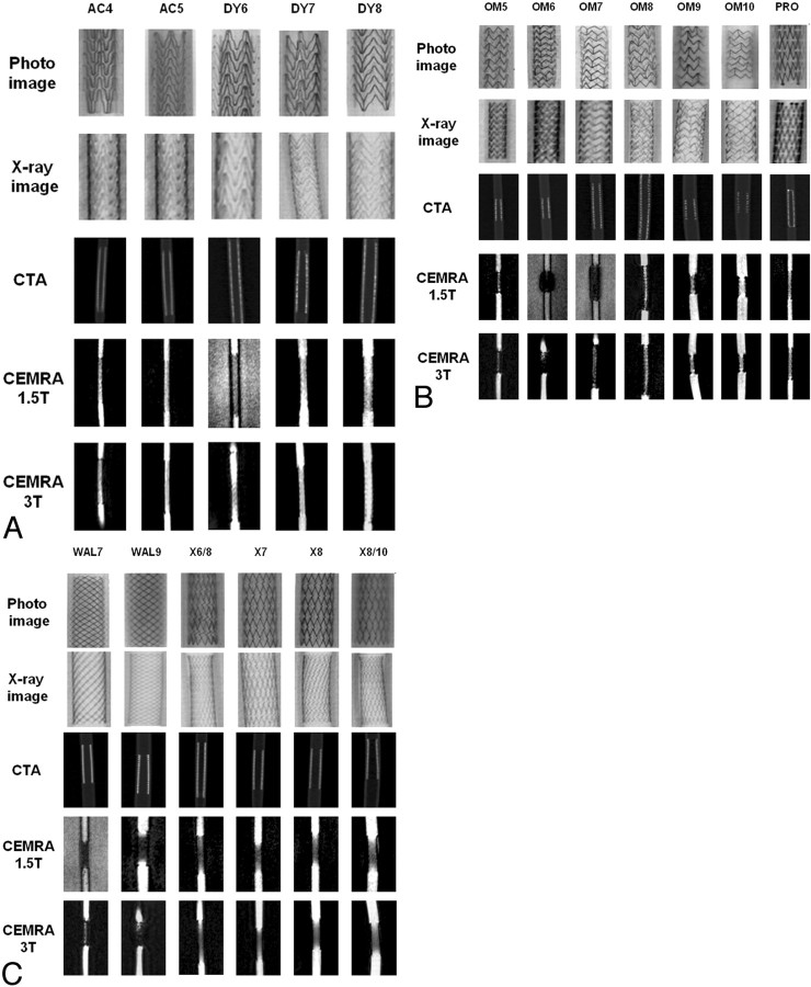
Background and purpose: CT angiography (CTA) and MR angiography (MRA) are increasingly used methods for evaluation of stented vessel segments. Our aim was to compare CTA, contrast-enhanced MRA (CE-MRA) at 1.5T, and CE-MRA at 3T for the visualization of carotid artery stents and to define the best noninvasive imaging technique as an alternative to conventional angiography for each stent.
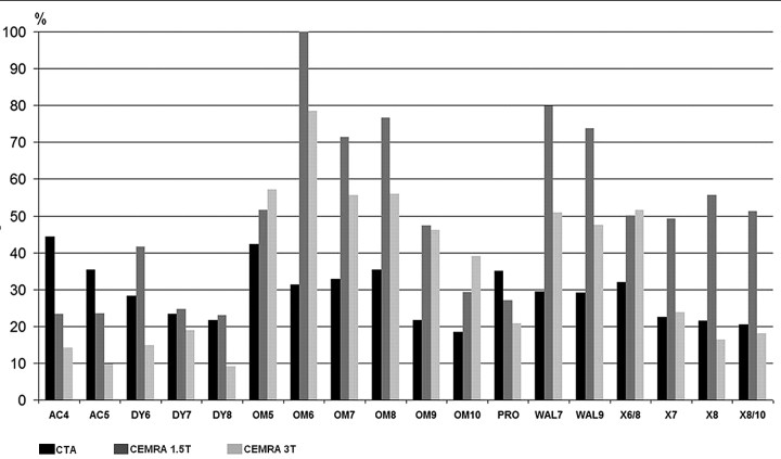
Materials and methods: CTA and CE-MRA appearances of 18 carotid artery stents of different designs and sizes (4.0 to 10.0 mm) were investigated in vitro. For each stent, artificial lumen narrowing (ALN) was calculated.
Results: With CE-MRA at 3T and at 1.5T, ALN in most nitinol stents was lower than that in the groups of stainless steel and cobalt alloy stents. In most nitinol stents and in both cobalt alloy stents, ALN was lower on CE-MRA at 3T than at 1.5T. In all stainless steel stents, ALN was lower on CTA than on CE-MRA. With CTA and CE-MRA, in most stents ALN decreased with increasing stent diameter.
Conclusions: CTA and CE-MRA evaluation of vessel patency after stent placement is possible but is considerably impaired by ALN. Investigators should be informed about the method of choice for every stent. Stent manufacturers should be aware of potential artifacts caused by their stents during noninvasive diagnostic methods such as CTA and CE-MRA.
Figures
Similar articles
-
In vitro comparison of different carotid artery stents: a pixel-by-pixel analysis using CT angiography and contrast-enhanced MR angiography at 1.5 and 3 T.Neuroradiology. 2010 Sep;52(9):823-30. doi: 10.1007/s00234-009-0625-5. Epub 2009 Nov 19. Neuroradiology. 2010. PMID: 19924409
-
3.0 Tesla contrast-enhanced MR angiography of carotid artery stents: in vitro measurements as compared with 1.5 Tesla.J Neuroradiol. 2006 Apr;33(2):75-80. doi: 10.1016/s0150-9861(06)77234-0. J Neuroradiol. 2006. PMID: 16733419
-
Carotid artery stents on CT angiography: in vitro comparison of different stent designs and sizes using 16-, 64- and 320-row CT scanners.J Neuroradiol. 2014 Oct;41(4):259-68. doi: 10.1016/j.neurad.2013.10.003. Epub 2014 Jan 9. J Neuroradiol. 2014. PMID: 24411522
-
Extracranial carotid MR imaging at 3T.Magn Reson Imaging Clin N Am. 2006 Feb;14(1):109-21. doi: 10.1016/j.mric.2005.12.003. Magn Reson Imaging Clin N Am. 2006. PMID: 16530639 Review.
-
Computed tomography angiography for the evaluation of carotid artery dissections.Front Neurol Neurosci. 2005;20:119-128. doi: 10.1159/000088156. Front Neurol Neurosci. 2005. PMID: 17290117 Review.
Cited by
-
Follow-up assessment of coiled intracranial aneurysms using zTE MRA as compared with TOF MRA: a preliminary image quality study.Eur Radiol. 2017 Oct;27(10):4271-4280. doi: 10.1007/s00330-017-4794-z. Epub 2017 Apr 5. Eur Radiol. 2017. PMID: 28382536
-
CE-MRA for follow-up of aneurysms post stent-assisted coiling.Interv Neuroradiol. 2012 Sep;18(3):275-83. doi: 10.1177/159101991201800305. Epub 2012 Sep 10. Interv Neuroradiol. 2012. PMID: 22958765 Free PMC article.
-
Optimization of MR Parameters of 3D TOF-MRA for Various Intracranial Stents at 3.0T MRI.Neurointervention. 2011 Aug;6(2):71-7. doi: 10.5469/neuroint.2011.6.2.71. Epub 2011 Aug 31. Neurointervention. 2011. PMID: 22125752 Free PMC article.
-
Angiographic CT: in vitro comparison of different carotid artery stents-does stent orientation matter?Neuroradiology. 2013 Jun;55(6):675-82. doi: 10.1007/s00234-013-1152-y. Epub 2013 Feb 15. Neuroradiology. 2013. PMID: 23411716
-
Comparing different MR angiography strategies of carotid stents in a vascular flow model: toward stent-specific recommendations in MR follow-up.Neuroradiology. 2011 May;53(5):359-65. doi: 10.1007/s00234-010-0753-y. Epub 2010 Aug 19. Neuroradiology. 2011. PMID: 20721544 Free PMC article.
References
-
- Eckstein HH, Ringleb P, Allenberg JR, et al. Results of the Stent-Protected Angioplasty versus Carotid Endarterectomy (SPACE) study to treat symptomatic stenoses at 2 years: a multinational, prospective, randomised trial. Lancet Neurol 2008;7:893–902 - PubMed
-
- Bendszus M, Koltzenburg M, Burger R, et al. Silent embolism in diagnostic cerebral angiography and neurointerventional procedures: a prospective study. Lancet 1999;354:1594–97 - PubMed
-
- Lenhart M, Volk M, Manke C, et al. Stent appearance at contrast-enhanced MR angiography: in vitro examination with 14 stents. Radiology 2000;217:173–78 - PubMed
-
- Wall A, Kugel H, Bachman R, et al. 3.0 T vs. 1.5 T MR angiography: in vitro comparison of intravascular stent artifacts. J Magn Reson Imaging 2005;22:772–79 - PubMed
Publication types
MeSH terms
Substances
LinkOut - more resources
Full Text Sources
Medical