Review
. 2010 Jan;31(1):15-23.
doi: 10.3174/ajnr.A1644. Epub 2009 Sep 12.

Neuroimaging and deep brain stimulation

Affiliations
Review

Neuroimaging and deep brain stimulation

D Dormont et al. AJNR Am J Neuroradiol. 2010 Jan.

Abstract

Deep brain stimulation (DBS) is a new neurosurgical method principally used for the treatment of Parkinson disease (PD). Many new applications of DBS are under development, including the treatment of intractable psychiatric diseases. Brain imaging is used for the selection of patients for DBS, to localize the target nucleus, to detect complications, and to evaluate the final electrode contact position. In patients with implanted DBS systems, there is a risk of electrode heating when MR imaging is performed. This contraindicates MR imaging unless specific precautions are taken. Involvement of neuroradiologists in DBS procedures is essential to optimize presurgical evaluation, targeting, and postoperative anatomic results. The precision of the neuroradiologic correlation with anatomic data and clinical outcomes in DBS promises to yield significant basic science and clinical advances in the future.


Figures

Fig 1.
Fig 1.
3D view of a postoperative MR acquisition in a patient with bilateral implantation of electrodes in the STN for the treatment of advanced PD. Caudate nuclei (blue), subthalamic nuclei (pink), electrodes (gray), and electrode contacts (blue) are segmented by using a 3D atlas described in Yelnik et al.. A, Anterior oblique view. B, Posterior oblique view. C, Zoom on the electrodes showing that their contacts are located inside the STN.
Fig 2.
Fig 2.
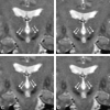
Coronal T2-weighted images showing the STN in a patient with advanced PD (spin-echo acquisition; TR/TE/NEX, 2200 ms/90 ms/2). Section thickness is 2 mm, located every 1 mm. A, The section is located 1 mm behind the anterior limit of the red nuclei. Both STNs are seen as almond-shaped hypointense structures above the locus niger (arrows). B, This section is located 1 mm in front of A, at the level of the anterior limit of the red nuclei. Both STNs are clearly seen (arrows). C and D, These sections are located 1 and 2 mm in front of B, showing the anterior extension of the STN (arrows).
Fig 3.
Fig 3.
Postoperative study of the position of electrode contacts in a patient with bilateral implantation of electrodes in the STN for the treatment of PD. Postoperative 3D MR imaging acquisition with fusion of anatomic data by using a histologically based deformable 3D atlas of the basal ganglia (Yelnik et al). A, 3D posterior oblique view shows the caudate nuclei (blue), the STN (pink), the locus niger (black), and the electrode contacts (yellow, active contacts; blue, inactive contacts; contacts inside or behind the STN are seen in the transparency). B, Frontal view through the active contact. The exact position of the contacts inside the metallic artifact on the MR imaging acquisition is indicated on each side by a yellow dot. Both contacts are located inside the STN (pink). Anatomic limits of the putamen (blue), caudate (blue), locus niger (black), thalamus (green), and optical tracts (orange) on the basis of the 3D atlas are shown. C, Axial view through the right active contact (yellow dot; the left active contact is above and is not seen in this view). The right active contact inside the STN (pink), caudate, putamen, and red nuclei (orange) can be seen. D, Right sagittal view through the right active contact (yellow dot). The same color code is used for caudate, thalamus, locus niger, and optical tract (orange).

References

    1. Benabid AL, Pollak P, Gervason C, et al. . Long-term suppression of tremor by chronic stimulation of the ventral intermediate thalamic nucleus. Lancet 1991; 337: 403–06 - PubMed
    1. Limousin P, Pollak P, Benazzouz A, et al. . Effect of parkinsonian signs and symptoms of bilateral subthalamic nucleus stimulation. Lancet 1995; 345: 91–95 - PubMed
    1. Landi A, Parolin M, Piolti R, et al. . Deep brain stimulation for the treatment of Parkinson's disease: the experience of the Neurosurgical Department in Monza. Neurol Sci 2003; 24: (suppl 1) S43–44 - PubMed
    1. Loher TJ, Burgunder JM, Pohle T, et al. . Long-term pallidal deep brain stimulation in patients with advanced Parkinson disease: 1-year follow-up study. J Neurosurg 2002; 96: 844–53 - PubMed
    1. Welter ML, Houeto JL, Tezenas du Montcel S, et al. . Clinical predictive factors of subthalamic stimulation in Parkinson's disease. Brain 2002; 125: 575–83 - PubMed