. 2009 Oct 15;461(7266):997-1001.
doi: 10.1038/nature08446. Epub 2009 Sep 13.

Spatiotemporal control of cell signalling using a light-switchable protein interaction

Affiliations

Spatiotemporal control of cell signalling using a light-switchable protein interaction

Anselm Levskaya et al. Nature. .

Abstract

Genetically encodable optical reporters, such as green fluorescent protein, have revolutionized the observation and measurement of cellular states. However, the inverse challenge of using light to control precisely cellular behaviour has only recently begun to be addressed; semi-synthetic chromophore-tethered receptors and naturally occurring channel rhodopsins have been used to perturb directly neuronal networks. The difficulty of engineering light-sensitive proteins remains a significant impediment to the optical control of most cell-biological processes. Here we demonstrate the use of a new genetically encoded light-control system based on an optimized, reversible protein-protein interaction from the phytochrome signalling network of Arabidopsis thaliana. Because protein-protein interactions are one of the most general currencies of cellular information, this system can, in principle, be generically used to control diverse functions. Here we show that this system can be used to translocate target proteins precisely and reversibly to the membrane with micrometre spatial resolution and at the second timescale. We show that light-gated translocation of the upstream activators of Rho-family GTPases, which control the actin cytoskeleton, can be used to precisely reshape and direct the cell morphology of mammalian cells. The light-gated protein-protein interaction that has been optimized here should be useful for the design of diverse light-programmable reagents, potentially enabling a new generation of perturbative, quantitative experiments in cell biology.


Conflict of interest statement

The authors declare no competing financial interests.

Figures

Figure 1
Figure 1
The phytochrome-PIF interaction can by used to reversibly translocate proteins to the plasma membrane in a light-controlled fashion. a, apoPhyB covalently binds to the chromophore phycocyanobilin (PCB) to form a light-sensitive holoprotein. PhyB undergoes conformational changes between the Pr and Pfr states catalyzed by red and infrared light, reversibly associating with the PIF domain only in the Pfr state. b, This heterodimerization interaction can be used to translocate a YFP-tagged PIF domain to PhyB tagged by mCherry and localized to the plasma membrane by the C-terminal caax motif of Kras. c, Phytochrome and PIF domains functional in mammalian cells were tested for reversible light-dependent recruitment of YFP to the plasma membrane using confocal microscopy. Previously published PIF constructs either failed to show visible recruitment or showed irreversible recruitment. Only PhyB constructs harboring tandem PAS repeats (unique to the plant phytochromes) showed detectable but reversible recruitment in vivo.
Figure 2
Figure 2
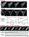
Confocal microscopy demonstrating the second-scale kinetics and photostability of the Phy-PIF photoswitchable membrane recruitment system. a, Confocal microscopy of NIH3T3 cells reveals rapid translocation of YFP between cytosol and plasma membrane under red and infrared light. Fitting exponentials to the cytoplasmic depletion of YFP gives typical time-constants of 1.3±0.1s for recruitment and 4±1s for dissociation (n=3). White rectangles show regions sampled for plotted traces. Arrows in graphs mark the timepoints shown. (Supplemental Movies 1,2) b, Rapid alternation between the 650 and 750 nM light can generate oscillations in the cytoplasmic concentrations of YFP. Absolute cytoplasmic concentration of YFP for this series is plotted along with the ratio change between time-points to adjust for photobleaching and cell-drift. The red and grey bars represent the standard deviations of the recruited and released cytosolic fluorescence, demonstrating near-fixed recruitment ratios over more than a hundred iterations. (Supplemental Movie 3) Scale bars 20μm.
Figure 3
Figure 3
Recruitment to the plasma membrane can be controlled spatially by simultaneously irradiating cells with patterned red and infra-red light. a, A nitrogen dye cell laser exciting a 650nm rhodamine dye was focused onto the sample plane of the microscope at 20Hz while IR-filtered white light continuously bathed the entire sample. b, A digital micromirror device focused onto the sample plane was used to send high-resolution patterns of 650nm/750nm light from a DG-4 source into the microscope under software control. This results in complementary red and infra-red distributions on the sample plane. c, TIRF imaging of localized membrane recruitment by a point source as in a shows highly localized YFP recruitment. (Supplementary movie 4) The recruited YFP spot's diameter is roughly 3μm and can be quickly moved by repositioning the laser. The final frame shows that the YFP spot is not merely bleed-through of the excitatory laser light, but genuine local fluorescent protein recruitment. d, TIRF movies of structured membrane recruitment by programmatically updating masks for red and infrared light by a digital micromirror device as in b were collected, revealing a faithful reproduction in the recruited YFP distribution of a movie of the cellular automaton ‘game-of-life glider’ that was projected (Supplementary movie 5). e, Images show the raw traces of titrated input 650nm light and recruited PIF-YFP. The plot at left shows the recruitment level as a function of 650nm ratio for three typical experiments. Inset shows the non-saturated regime. Scale bars 20μm.
Figure 4
Figure 4
Rho-family G-protein signalling can be controlled by the light-activated translocation system. a, The catalytic DH-PH domains of RhoGEFs Tiam and Intersectin activate their respective G-proteins Rac1 and Cdc42 which in turn act through effector proteins to modify the actin cytoskeleton. b, Recruitee constructs with Tiam DH-PH domains were assayed for their ability to induce lamellipodia in NIH3T3 by exposing serum-depleted cells transfected with the indicated constructs to red (650nm) light and counting the percentage of cells that produced lamellipodia within 20min under live microscopy. Error bars s.e.m., (n=2, avg. 30 cells; p-value=.0004 for Tiam) c, Local induction and ‘extrusion’ of lamellipodia in live NIH3T3 cells was demonstrated by globally irradiating the whole sample with a infrared (750nm) light source while focusing a red (650nm) laser onto a small portion of the cell as in 2a and slowly extending this red-targeted region from the cell body. Superimposed outlines of the cell show directed extension 30μm along the line of light movement. (Supplemental Movie 7) d, Cdc42-GTP binding domain (WASP-GBD) linked to mCherry was used to measure the “response function” of Intersectin DHPH recruitment over several iterations in time and in space at equilibrium. (Supplemental Movie 11) Scale bars 20μm.

References

    1. Szobota S, et al. Remote control of neuronal activity with a light-gated glutamate receptor. Neuron. 2007;54:535–45. - PubMed
    1. Boyden ES, Zhang F, Bamberg E, Nagel G, Deisseroth K. Millisecond-timescale, genetically targeted optical control of neural activity. Nat Neurosci. 2005;8:1263–8. - PubMed
    1. Han X, Boyden ES. Multiple-color optical activation, silencing, and desynchronization of neural activity, with single-spike temporal resolution. PLoS ONE. 2007;2:e299. - PMC - PubMed
    1. Mettetal JT, Muzzey D, Gómez-Uribe C, van Oudenaarden A. The frequency dependence of osmo-adaptation in Saccharomyces cerevisiae. Science. 2008;319:482–4. - PMC - PubMed
    1. Bennett MR, et al. Metabolic gene regulation in a dynamically changing environment. Nature. 2008;454:1119–22. - PMC - PubMed

Publication types

MeSH terms

Substances