. 2009 Oct;15(10):1215-8.
doi: 10.1038/nm.2025. Epub 2009 Sep 13.

Molecular signatures of disease brain endothelia provide new sites for CNS-directed enzyme therapy

Affiliations

Molecular signatures of disease brain endothelia provide new sites for CNS-directed enzyme therapy

Yong Hong Chen et al. Nat Med. 2009 Oct.

Abstract

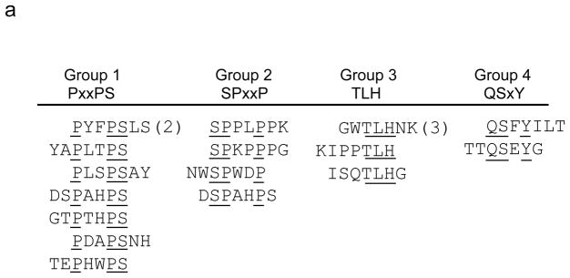
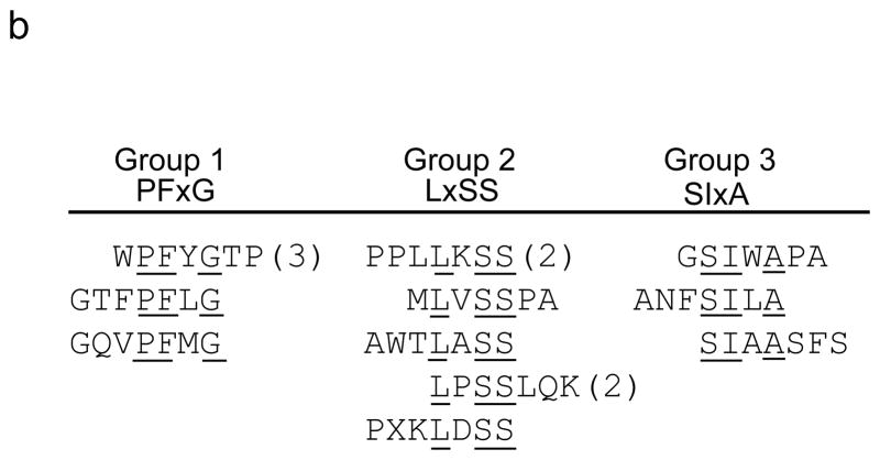
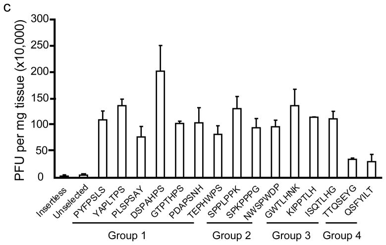
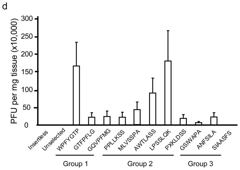
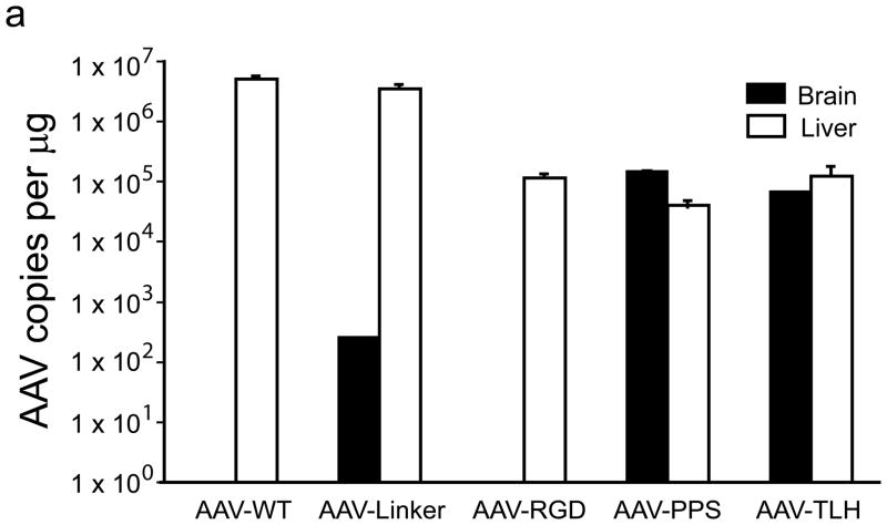
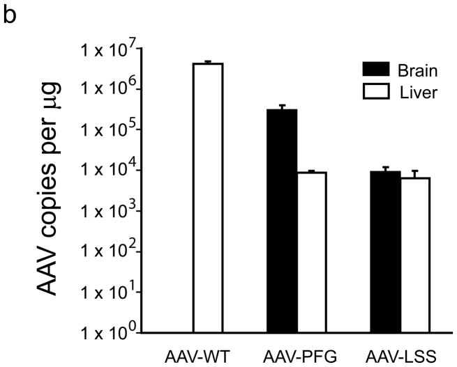
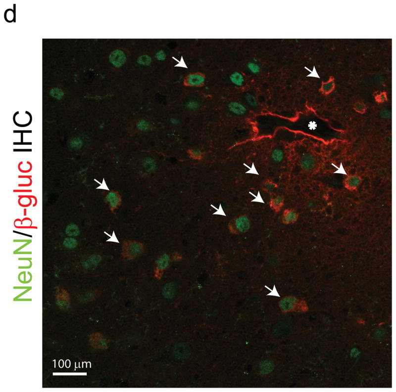
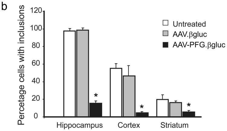
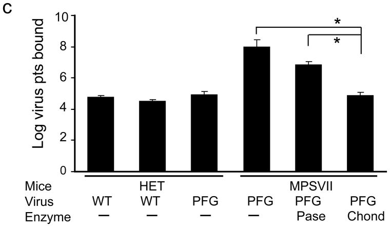
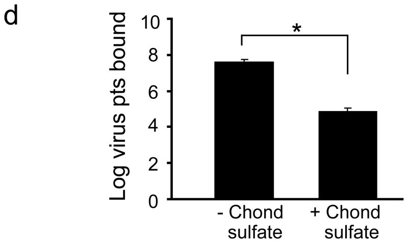
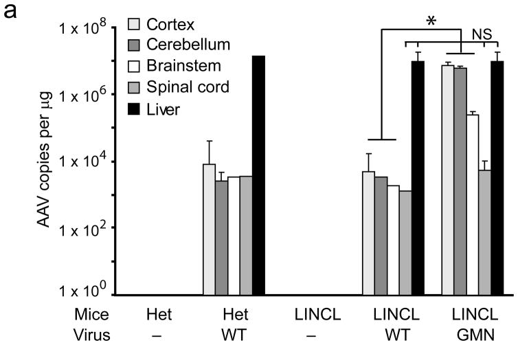
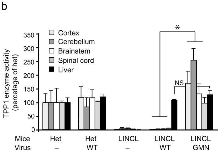
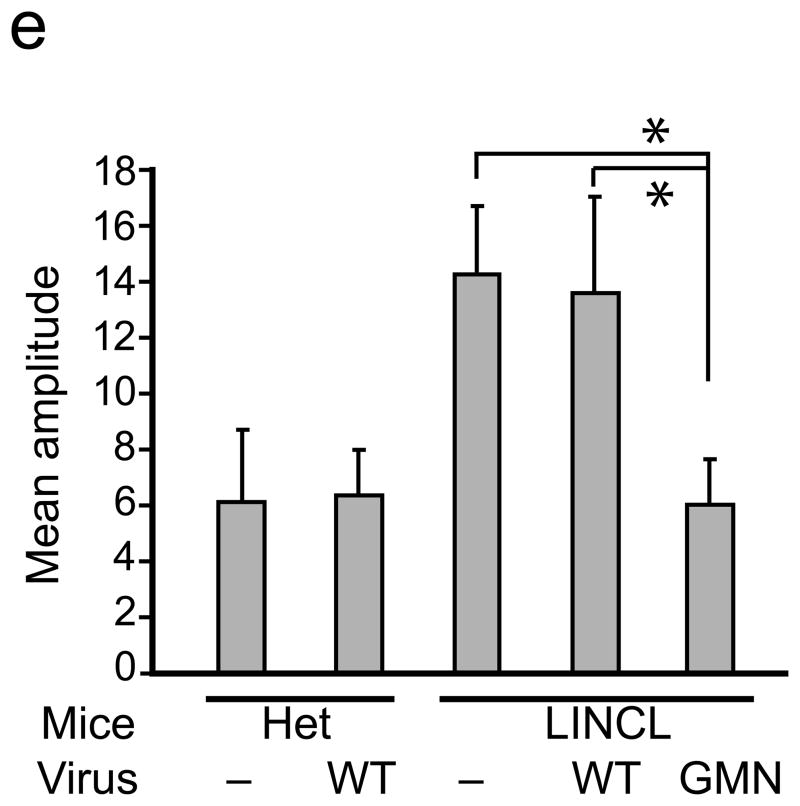
The brain vasculature forms an immense network such that most neural cells are in contact with a microvessel. Here we tested the hypothesis that endothelia lining these vessels can be harnessed to create a cellular reservoir of enzyme replacement therapy to diseased brain. As a model system, we used mice with central nervous system (CNS) deficits due to lysosomal storage disease (LSD mice). The basic premise of this work is that recombinant enzyme expressed in, and secreted from, the vascular endothelia will be endocytosed by underlying neurons and glia, decreasing neuropathology. We screened a phage library in vivo by panning to identify peptides that bound the vascular endothelia in diseased and wild-type mice. Epitopes binding diseased brain were distinct from those panned from normal brain. Moreover, different epitopes were identified in two distinct LSD disease models, implying a unique vascular signature imparted by the disease state. Presentation of these epitopes on the capsid of adeno-associated virus (AAV) expanded the biodistribution of intravenously injected AAV from predominantly liver to include the CNS. Peripheral injection of the epitope-modified AAVs expressing the enzymes lacking in LSD mice reconstituted enzyme activity throughout the brain and improved disease phenotypes in two distinct disease models.


Figures

Figure 1
Figure 1
In vivo phage display panning to identify peptide motifs with high affinity for cerebral vasculature. (a,b) After in vivo phage display panning, phage with distinct peptide motifs were identified from wildtype (a) and MPS VII (b) mice. (c,d) Purified selected phage were individually injected via tail vein into naïve mice to validate enrichment in brain. Data show phage titers recovered from the cerebral vasculature of heterozygous (c) and MPS VII (d) mice. Data presented as mean ± SEM. One-way ANOVA revealed group (n=3 all groups) as a significant factor for both Het (F2,50 = 20; p<0.0001) and MPS VII (F2,38 = 251.7; p<0.0001) mice. For Het mice, 13 of 15 epitopes were significantly enriched in brain relative to mice injected with the unselected library. (Groups 1–3, p<0.05, Dunnett’s post hoc). For MPS VII mice, 9 of 11 epitopes were significantly enriched in brain relative to mice injected with the unselected library (for all but GSWAPA and SSIAASFS, p<0.05, Dunnett’s post hoc).
Figure 1
Figure 1
In vivo phage display panning to identify peptide motifs with high affinity for cerebral vasculature. (a,b) After in vivo phage display panning, phage with distinct peptide motifs were identified from wildtype (a) and MPS VII (b) mice. (c,d) Purified selected phage were individually injected via tail vein into naïve mice to validate enrichment in brain. Data show phage titers recovered from the cerebral vasculature of heterozygous (c) and MPS VII (d) mice. Data presented as mean ± SEM. One-way ANOVA revealed group (n=3 all groups) as a significant factor for both Het (F2,50 = 20; p<0.0001) and MPS VII (F2,38 = 251.7; p<0.0001) mice. For Het mice, 13 of 15 epitopes were significantly enriched in brain relative to mice injected with the unselected library. (Groups 1–3, p<0.05, Dunnett’s post hoc). For MPS VII mice, 9 of 11 epitopes were significantly enriched in brain relative to mice injected with the unselected library (for all but GSWAPA and SSIAASFS, p<0.05, Dunnett’s post hoc).
Figure 2
Figure 2
Peptide epitopes expand the tropism of AAV2. (a,b) Viral genomes in brain and liver of wildtype (a) and MPS VII (b) mice as quantified by Q-PCR after peripheral injection. Data are mean ± SEM. (a) AAV-PPS and AAV-TLH are significantly enriched in brain (p<0.001, students t-test) vs. AAV-WT. (b) AAV-PFG and AAV-LSS are significantly enriched in brain over AAV-WT (p<0.001, student’s t-test). (c) AAV-PFG.βgluc transduces the cerebral vasculature of MPS VII mice after tail vein injection. Representative sections were stained for β-glucuronidase activity in situ, which leaves a red precipitate. Representative photomicrographs from sections show that vessels throughout the brain display activity when MPS VII mice (left and middle panels), but not wildtype mice (upper right), were injected. (d) Confocal microscopy of sections stained with anti-β-glucuronidase (red) and anti-NeuN (green) reveals vascular and neuronal β-glucuronidase staining, respectively (denoted by arrows).
Figure 2
Figure 2
Peptide epitopes expand the tropism of AAV2. (a,b) Viral genomes in brain and liver of wildtype (a) and MPS VII (b) mice as quantified by Q-PCR after peripheral injection. Data are mean ± SEM. (a) AAV-PPS and AAV-TLH are significantly enriched in brain (p<0.001, students t-test) vs. AAV-WT. (b) AAV-PFG and AAV-LSS are significantly enriched in brain over AAV-WT (p<0.001, student’s t-test). (c) AAV-PFG.βgluc transduces the cerebral vasculature of MPS VII mice after tail vein injection. Representative sections were stained for β-glucuronidase activity in situ, which leaves a red precipitate. Representative photomicrographs from sections show that vessels throughout the brain display activity when MPS VII mice (left and middle panels), but not wildtype mice (upper right), were injected. (d) Confocal microscopy of sections stained with anti-β-glucuronidase (red) and anti-NeuN (green) reveals vascular and neuronal β-glucuronidase staining, respectively (denoted by arrows).
Figure 3
Figure 3
Intravenous delivery of epitope-modified virus improves neuropathology in MPS VII mice. (a) Representative sections of cerebral cortex, hippocampus, striatum and cerebellum of MPS VII mice harvested after tail vein injection with either AAV-WT.βluc (left panels) or AAV-PFG.βluc (right panels) expressing β-glucuronidase. Yellow asterisk, denotes region magnified in inset (lower right, all panels) for better visualization of storage vacuoles. Arrows in inset point to vacuoles. Scale bar = 50 μm for all panels. (b) Quantitation of vacuolar storage in various brain regions. Tail vein injection of AAV-PFG.βluc but not AAV-WT.βluc significantly reduced lysosomal storage vacuoles in hippocampus, cortex and striatum (*p<0.001, Tukeys post hoc). (c) Binding of AAV-PFG to cerebral vasculature requires chondroitin sulfate. Purified brain microvessels from heterozygous or MPS VII mice were incubated with the reagents indicated, and bound viral particles quantified. Data presented as mean ± SEM. (*,p<0.001, Dunnett’s post hoc). (d) Binding of AAV-PFG to purified brain vasculature from MPS VII mice in the presence or absence of chondroitin sulfate. Data presented as mean ± SEM. *<0.01, student’s t-test.
Figure 3
Figure 3
Intravenous delivery of epitope-modified virus improves neuropathology in MPS VII mice. (a) Representative sections of cerebral cortex, hippocampus, striatum and cerebellum of MPS VII mice harvested after tail vein injection with either AAV-WT.βluc (left panels) or AAV-PFG.βluc (right panels) expressing β-glucuronidase. Yellow asterisk, denotes region magnified in inset (lower right, all panels) for better visualization of storage vacuoles. Arrows in inset point to vacuoles. Scale bar = 50 μm for all panels. (b) Quantitation of vacuolar storage in various brain regions. Tail vein injection of AAV-PFG.βluc but not AAV-WT.βluc significantly reduced lysosomal storage vacuoles in hippocampus, cortex and striatum (*p<0.001, Tukeys post hoc). (c) Binding of AAV-PFG to cerebral vasculature requires chondroitin sulfate. Purified brain microvessels from heterozygous or MPS VII mice were incubated with the reagents indicated, and bound viral particles quantified. Data presented as mean ± SEM. (*,p<0.001, Dunnett’s post hoc). (d) Binding of AAV-PFG to purified brain vasculature from MPS VII mice in the presence or absence of chondroitin sulfate. Data presented as mean ± SEM. *<0.01, student’s t-test.
Figure 4
Figure 4
An epitope panned from TPP-1 deficient mice extends AAV tropism to brain and allows correction of CNS deficits. (a)AAV-GMN vectors were significantly enriched in cortex, cerebellum and brainstem after tail vein injection, in contrast to those same regions harvested from LINCL injected with AAV-WT vectors (*,p<0.005). Vector genomes in spinal cord and liver were similar between the two groups (p=0.12 and 0.61, respectively; student’s t-test). NS, not significant. (b) Six weeks after tail vein injection, enzyme activity was significantly higher in all brain regions from AAV-GMN.TPP1 vs. AAV-WT.TPP1 treated LINCL mice (*, p<0.005; students t-test). (c) AAV-GMN.TPP1 but not AAV-WT.TPP1 improves glial activation in LINCL motor cortex. Representative photomicrographs of heterozygous mice or LINCL mice treated with AAV-WT.TPP1 (upper and middle panels) demonstrate the extent of glial activation in the model at P90. LINCL mice treated with AAV-GMN.TPP1 show dramatically reduced GFAP immunoreactivity. Scale bar = 100 μm. Quantitation using Image J indicates significant effects (*, p<0.001, Tukeys post hoc). Het vs. AAV-GMN-treated LINCL mice were not significantly different. (d) Tail vein injection of AAV-GMN.TPP1 prevents loss of deep cerebellar nuclei, in contrast to AAV-WT.TPP1. Scale bar = 500 μm. (e) AAV-GMN.TPP1, but not AAV-WT.TPP1 significantly improves the tremor phenotype in LINCL mice (* p<0.001, Dunnett’s post hoc). Tail vein injection of AAV-WT.TPP1 had no affect on the tremor phenotype of LINCL mice.
Figure 4
Figure 4
An epitope panned from TPP-1 deficient mice extends AAV tropism to brain and allows correction of CNS deficits. (a)AAV-GMN vectors were significantly enriched in cortex, cerebellum and brainstem after tail vein injection, in contrast to those same regions harvested from LINCL injected with AAV-WT vectors (*,p<0.005). Vector genomes in spinal cord and liver were similar between the two groups (p=0.12 and 0.61, respectively; student’s t-test). NS, not significant. (b) Six weeks after tail vein injection, enzyme activity was significantly higher in all brain regions from AAV-GMN.TPP1 vs. AAV-WT.TPP1 treated LINCL mice (*, p<0.005; students t-test). (c) AAV-GMN.TPP1 but not AAV-WT.TPP1 improves glial activation in LINCL motor cortex. Representative photomicrographs of heterozygous mice or LINCL mice treated with AAV-WT.TPP1 (upper and middle panels) demonstrate the extent of glial activation in the model at P90. LINCL mice treated with AAV-GMN.TPP1 show dramatically reduced GFAP immunoreactivity. Scale bar = 100 μm. Quantitation using Image J indicates significant effects (*, p<0.001, Tukeys post hoc). Het vs. AAV-GMN-treated LINCL mice were not significantly different. (d) Tail vein injection of AAV-GMN.TPP1 prevents loss of deep cerebellar nuclei, in contrast to AAV-WT.TPP1. Scale bar = 500 μm. (e) AAV-GMN.TPP1, but not AAV-WT.TPP1 significantly improves the tremor phenotype in LINCL mice (* p<0.001, Dunnett’s post hoc). Tail vein injection of AAV-WT.TPP1 had no affect on the tremor phenotype of LINCL mice.

Comment in

  • Gateway to the diseased brain.
    Mingozzi F, High KA. Mingozzi F, et al. Nat Med. 2009 Oct;15(10):1123-4. doi: 10.1038/nm1009-1123. Nat Med. 2009. PMID: 19812565 No abstract available.

References

    1. Work LM, et al. Vascular bed-targeted in vivo gene delivery using tropism-modified adeno-associated viruses. Mol Ther. 2006;13:683–693. - PubMed
    1. Grifman M, et al. Incorporation of tumor-targeting peptides into recombinant adeno-associated virus capsids. Molecular Therapy. 2001;3(6):964–975. - PubMed
    1. Muller OJ, et al. Random peptide libraries displayed on adeno-associated virus to select for targeted gene therapy vectors. Nat Biotechnol. 2003;21:1040–1046. - PubMed
    1. Perabo L, et al. Heparan sulfate proteoglycan binding properties of adeno-associated virus retargeting mutants and consequences for their in vivo tropism. J Virol. 2006;80:7265–7269. - PMC - PubMed
    1. Vogler C, et al. A novel model of murine mucopolysaccharidosis type VII due to an intracisternal a particle element transposition into the beta-glucuronidase gene: clinical and pathologic findings. Pediatr Res. 2001;49:342–348. - PubMed

Publication types