. 2009 Sep 14;4(9):e7030.
doi: 10.1371/journal.pone.0007030.

Identification of gemin5 as a novel 7-methylguanosine cap-binding protein

Affiliations

Identification of gemin5 as a novel 7-methylguanosine cap-binding protein

Shelton S Bradrick et al. PLoS One. .

Abstract

Background: A unique attribute of RNA molecules synthesized by RNA polymerase II is the presence of a 7-methylguanosine (m(7)G) cap structure added co-transcriptionally to the 5' end. Through its association with trans-acting effector proteins, the m(7)G cap participates in multiple aspects of RNA metabolism including localization, translation and decay. However, at present relatively few eukaryotic proteins have been identified as factors capable of direct association with m(7)G.

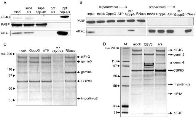
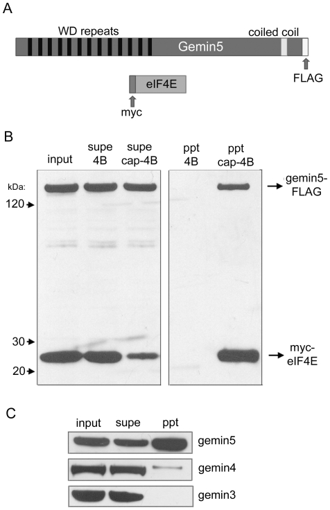
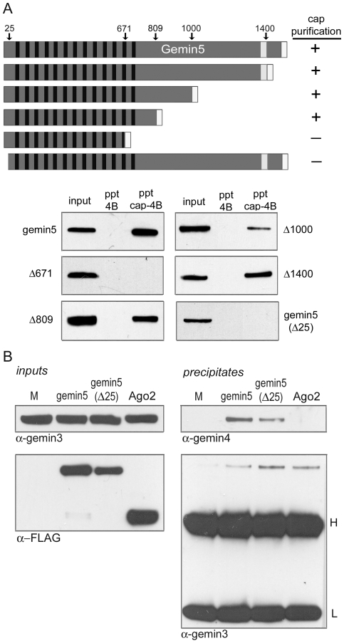
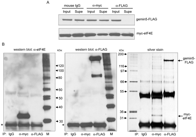
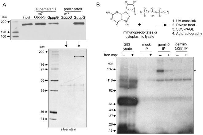
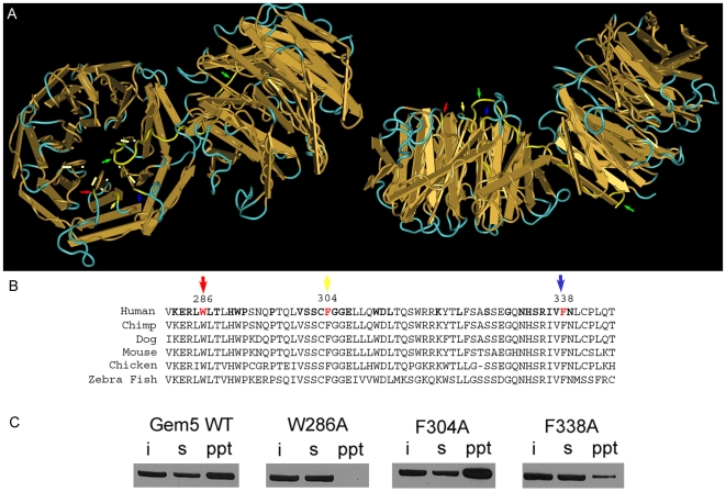
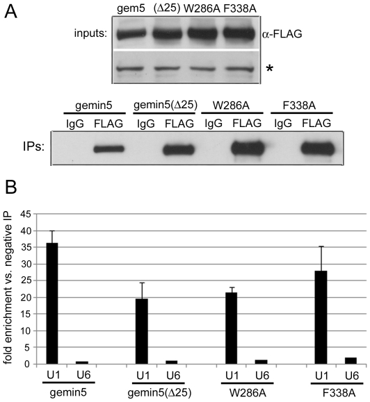
Methodology/principal findings: Employing an unbiased proteomic approach, we identified gemin5, a component of the survival of motor neuron (SMN) complex, as a factor capable of direct and specific interaction with the m(7)G cap. Gemin5 was readily purified by cap-affinity chromatography in contrast to other SMN complex proteins. Investigating the underlying basis for this observation, we found that purified gemin5 associates with m(7)G-linked sepharose in the absence of detectable eIF4E, and specifically crosslinks to radiolabeled cap structure after UV irradiation. Deletion analysis revealed that an intact set of WD repeat domains located in the N-terminal half of gemin5 are required for cap-binding. Moreover, using structural modeling and site-directed mutagenesis, we identified two proximal aromatic residues located within the WD repeat region that significantly impact m(7)G association.

Conclusions/significance: This study rigorously identifies gemin5 as a novel cap-binding protein and describes an unprecedented role for WD repeat domains in m(7)G recognition. The findings presented here will facilitate understanding of gemin5's role in the metabolism of non-coding snRNAs and perhaps other RNA pol II transcripts.


Conflict of interest statement

Competing Interests: The authors have declared that no competing interests exist.

Figures

Figure 1
Figure 1. Characterization of proteins purified by cap-affinity chromatography.
(A) HeLa cytoplasmic lysate was applied to m7G-sepharose 4B or sepharose 4B alone. Precipitates (ppt) were washed extensively and then resuspended in sample buffer. Levels of eIF4G, PABP and eIF4E in input and supernatant (supe) samples were examined by western blot. Input samples represent 10% of total. (B) Cap-affinity chromatography was performed in the presence of the indicated nucleotide (0.1 mM final concentration) or 0.1 mg/ml RNase A and PABP/eIF4E were detected by western blot. (C) The same precipitate samples shown in (B) were subjected to silver stain detection. The identities of proteins determined by mass spectrometry are indicated. (D) HeLa lysates were derived from normally growing cells (mock), cells infected with CBV3 at four hours post-infection, or cells acutely stressed with 0.1 mM arsenite for 30 minutes. Proteins precipitated with cap-sepharose were then analyzed by silver stain.
Figure 2
Figure 2. Gemin5 isolated by cap-affinity is not bound to the SMN complex.
(A) Schematic representation of gemin5 and eIF4E indicating locations of WD repeats, the putative coiled coil domain, and epitope tags. (B) 293T cells were co-transfected with expression constructs encoding FLAG-tagged gemin5 and myc-tagged eIF4E. Cell lysate was derived 24 hours later and applied to cap-sepharose 4B or sepharose 4B. Tagged proteins were detected with α-FLAG and α-myc antibodies. (C) Purification of proteins by cap-affinity was performed as in figure 1A and levels of endogenous gemin3–5 were examined in input, supernatant and precipitate samples.
Figure 3
Figure 3. Mapping of determinants within gemin5 required for association with m7G-sepharose.
(A) Multiple C-terminal truncations and a single N-terminal truncation of FLAG-tagged gemin5 were evaluated by cap-affinity chromatography. Deletion sites within gemin5 are indicated above by amino acid number. (B) FLAG-tagged gemin5, gemin5(Δ25) variant, and Ago2 expression constructs were transfected into 293T cells along with pcDNA3 alone (M). Input samples used for co-IP assays were analyzed by detection of gemin3 and over-expressed FLAG-tagged proteins (left). Each lysate was subjected to IP using α-FLAG antibody and precipitate samples were analyzed for the presence of gemin3 and gemin4. Positions of heavy (H) and light (L) antibody chains are indicated.
Figure 4
Figure 4. Gemin5 fails to detectably interact with eIF4E in co-IP experiments.
293T cells were co-transfected with gemin5 and eIF4E expression constructs as in figure 2 and cytoplasmic lysate was used for IP using the indicated murine antibodies (IgG, α-myc, and α-FLAG). (A) Input and supernatant samples assayed for levels of over-expressed proteins. (B) Each IP was analyzed by western blot with α-eIF4E (left) and α-FLAG (middle) rabbit antibodies, and by silver stain (right). Blots were intentionally over-exposed to assess possible co-IP. Asterisks indicate mouse light antibody chain used in IP reactions that cross reacts with rabbit secondary antibody.
Figure 5
Figure 5. Gemin5 binds directly to the m7G cap structure.
(A) FLAG-tagged gemin5 was immunoprecipitated and released from the resin with FLAG peptide. Eluted protein was subsequently applied to cap-sepharose in the presence of free m7GpppG or GpppG competitors. Precipitation of FLAG-gemin5 was monitored by western blot and silver stain analysis. (B) A representation of the m7G cap structure is shown with position of radiolabeled α-phosphate indicated by an asterisk. A short RNA transcript was synthesized and then modified with an m7G cap using [α-32P]-GTP and guanylyltransferase (see experimental procedures for details). Purified capped RNA was incubated in the presence or absence 0.1 mM free cap analog with 1) 293T cytoplasmic lysate, 2) eluate from α-FLAG IP of lysate from pcDNA3-transfected cells (mock IP), or 3) FLAG-tagged gemin5 or gemin5(Δ25) immunopurified as in (A). Binding reactions were irradiated with UV light, incubated with RNase cocktail, and then analyzed by SDS-PAGE followed by autoradiography as indicated.
Figure 6
Figure 6. Identification of amino acid residues that affect m7G recognition by gemin5.
(A) PHYRE analysis was used to model the gemin5 WD repeat domains onto the structure of actin-interacting protein 1 (AIP-1; see Materials and Methods). The backbone of the AIP-1 structure, consisting of two β-propellers, is shown with indicated locations of amino acids highlighted in yellow. A 90° x-axis rotation of the left structure is shown at right. Arrows indicate positions of W286 (red), F304 (yellow), F338 (blue) and the N-terminal 25 amino acids of AIP-1 (green). Note that the N-terminus of AIP-1 forms a β-sheet in the last WD repeat domain of the second β-propeller before looping into the first β-propeller to form another β-sheet. PHYRE analysis predicts only the second β-sheet in gemin5. AIP-1 structures were visualized using Cn3D . (B) Alignments of gemin5 sequences from selected vertebrate species is shown. Bold letters in the human sequence indicate uniform conservation and positions of residues 286, 304 and 338 are indicated. (C) Cap-binding assays were performed with wild-type gemin5-FLAG and variants as in previous figures. Input (i), supernatant (s), and precipitate (ppt) samples are indicated.
Figure 7
Figure 7. Gemin5 mutations that affect m7G interaction reduce association with U1 snRNA.
(A) Gemin5-FLAG and the indicated variants were transiently expressed in 293T cells and then immunoprecipitated with α-FLAG antibody or negative control mouse IgG. A 10% fraction of IP samples along with input samples were subjected to α-FLAG western blot. The asterisk indicates a cross-reactive band that serves as a loading control. The remaining IPs were used for RNA extraction. (B) RT-qPCR was performed on extracted RNAs for measurements of U1 and U6 snRNA levels in positive and negative IP samples for each gemin5 variant. Error bars indicate values for standard deviation.

References

    1. Shuman S. Structure, mechanism, and evolution of the mRNA capping apparatus. Prog Nucleic Acid Res Mol Biol. 2001;66:1–40. - PubMed
    1. Saha N, Schwer B, Shuman S. Characterization of human, Schizosaccharomyces pombe, and Candida albicans mRNA cap methyltransferases and complete replacement of the yeast capping apparatus by mammalian enzymes. J Biol Chem. 1999;274:16553–16562. - PubMed
    1. Konarska MM, Padgett RA, Sharp PA. Recognition of cap structure in splicing in vitro of mRNA precursors. Cell. 1984;38:731–736. - PubMed
    1. Proudfoot NJ, Furger A, Dye MJ. Integrating mRNA processing with transcription. Cell. 2002;108:501–512. - PubMed
    1. Wilusz CJ, Wormington M, Peltz SW. The cap-to-tail guide to mRNA turnover. Nat Rev Mol Cell Biol. 2001;2:237–246. - PubMed

Publication types