. 2009 Sep 14;4(9):e6989.
doi: 10.1371/journal.pone.0006989.

O antigen allows B. parapertussis to evade B. pertussis vaccine-induced immunity by blocking binding and functions of cross-reactive antibodies

Affiliations

O antigen allows B. parapertussis to evade B. pertussis vaccine-induced immunity by blocking binding and functions of cross-reactive antibodies

Xuqing Zhang et al. PLoS One. .

Abstract

Although the prevalence of Bordetella parapertussis varies dramatically among studies in different populations with different vaccination regimens, there is broad agreement that whooping cough vaccines, composed only of B. pertussis antigens, provide little if any protection against B. parapertussis. In C57BL/6 mice, a B. pertussis whole-cell vaccine (wP) provided modest protection against B. parapertussis, which was dependent on IFN-gamma. The wP was much more protective against an isogenic B. parapertussis strain lacking O-antigen than its wild-type counterpart. O-antigen inhibited binding of wP-induced antibodies to B. parapertussis, as well as antibody-mediated opsonophagocytosis in vitro and clearance in vivo. aP-induced antibodies also bound better in vitro to the O-antigen mutant than to wild-type B. parapertussis, but aP failed to confer protection against wild-type or O antigen-deficient B. parapertussis in mice. Interestingly, B. parapertussis-specific antibodies provided in addition to either wP or aP were sufficient to very rapidly reduce B. parapertussis numbers in mouse lungs. This study identifies a mechanism by which one pathogen escapes immunity induced by vaccination against a closely related pathogen and may explain why B. parapertussis prevalence varies substantially between populations with different vaccination strategies.


Conflict of interest statement

Competing Interests: The authors have declared that no competing interests exist.

Figures

Figure 1
Figure 1. B. parapertussis is more susceptible to wP–induced immunity in the absence of O-antigen.
Groups of four naïve (black) or wP vaccinated (white) C57BL/6 mice were challenged with the indicated bacteria. (A) The number of CFUs recovered from the respiratory tract on day 3 post-challenge is expressed as the Log10 mean ± the standard error. Decreases in Log10CFU in vaccinated mice compared to naïve mice on day 3 post-challenge are indicated underneath the x axes. (B) The change in CFU number over the first 3 days after challenge is expressed as change in Log10 mean ± the standard error. * indicates P≤0.05. ** indicates P≤0.01. The limit of detection is indicated as the lower limit of the y axes.
Figure 2
Figure 2. aP does not reduce B. parapertussis numbers.
Groups of four C57BL/6 mice were vaccinated with adjuvant only (black) or aP (white), challenged with the indicated bacteria and sacrificed on day 3 post-challenge. The numbers of CFUs recovered from the respiratory tract are expressed as the Log10 mean ± the standard error. ** indicates P≤0.01. The limit of detection is indicated as the lower limit of the y axes.
Figure 3
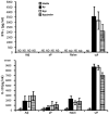
Figure 3. Splenic production of IFN-γ and IL-10 is cross-reactive.
Splenocytes from groups of four naïve C57BL/6 mice or mice vaccinated with the indicated vaccine were stimulated with media only (vertically hatched), heat-killed B. pertussis (black), B. parapertussis (white) or O-antigen deficient B. parapertussis (horizontally hatched) for 3 days. The concentration of IFN-γ and IL-10 in the supernatant is expressed as the mean ± the standard error. ND indicates none detected.
Figure 4
Figure 4. IFN-γ contributes to the protection against B. parapertussis by wP.
Groups of four naïve (black and horizontally hatched) or wP vaccinated (white and vertically hatched) C57BL/6 mice were untreated (−) (black and white) or i.p. injected with (+) (horizontally and vertically hatched) anti-IFN-γ antibody, challenged with B. parapertussis and sacrificed 3 days post-challenge. The number of CFUs throughout the respiratory tract is expressed as the Log10 mean ± the standard error. * indicates P≤0.05. ** indicates P≤0.01. The limit of detection is indicated as the lower limit of the y axes.
Figure 5
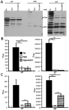
Figure 5. O-antigen inhibits the binding of B. pertussis vaccine-induced antibodies to live, but not denatured, B. parapertussis cells.
(A) Western blots were performed on lysates of indicated bacteria probed with serum antibodies collected from mice that were vaccinated with aP, adjuvant only (Adj) or wP. (B) Heat-inactivated or (C) live B. pertussis (black), B. parapertussis (white) or O-antigen deficient B. parapertussis (hatched) were coated on ELISA plate. Serum antibody titer of mice vaccinated with aP or wP is expressed as mean of the end point titers of four independent samples ± the standard error. ** indicates P≤0.01. The dashed line indicates the limit of detection.
Figure 6
Figure 6. O-antigen decreases the opsonization of B. parapertussis by B. pertussis vaccine-induced antibodies.
Representative histograms show flow cytometric analysis of indicated bacteria opsonized with naïve serum (white) or (A) aP, (B) wP, (C) wPP-induced serum (grey) and stained with RPE-labeled goat F(ab)2 fragments of anti-mouse IgG. (D) Mean red-fluorescence of B. pertussis (black), B. parapertussis (white), O-antigen deficient B. parapertussis (hatched) opsonized with indicated serum from four individual mice ± the standard error is shown. AU indicates arbitrary units. ** indicates P≤0.01.
Figure 7
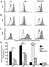
Figure 7. O-antigen blocks B. pertussis vaccine-induced antibodies from mediating adherence of B. parapertussis to PMNs.
Naïve serum (white) or (A) aP, (B) wP, (C) wPP-induced serum (grey)-opsonized GFP-expressing bacteria were incubated with freshly isolated human peripheral blood PMNs. Representative histograms of flow cytometry analysis of these cells are shown. (D) Mean green fluorescence associated with PMNs incubated with GFP-expressing B. pertussis (black), B. parapertussis (white) or O-antigen deficient B. parapertussis (hatched) opsonized with four independent indicated serum ± the standard error is shown. AU indicates arbitrary units. ** indicates P≤0.01.
Figure 8
Figure 8. O-antigen blocks B. pertussis vaccine-induced, antibody-mediated phagocytosis of B. parapertussis.
Freshly isolated human peripheral blood PMNs were incubated for 20 min or 1 h and 20 min with GFP-expressing B. pertussis (black), B. parapertussis (white), or O-antigen deficient B. parapertussis (hatched), that are opsonized with serum from aP, wP, wPP-vaccinated or naïve mice. The cell surface bound bacteria were detected by incubation with RPE-labeled goat F(ab')2 fragments of anti-mouse IgG. Mean phagocytosis calculated from the decrease in red-fluorescence of green-positive cells incubated for 1 h and 20 min compared to that incubated for 20 min of experiments done with 4 independent serum samples ± the standard error was shown. AU indicates arbitrary units. ** indicates P≤0.01.
Figure 9
Figure 9. Passive transfer of wP–induced serum antibodies mediates clearance of O-antigen deficient, but not wild-type, B. parapertussis from mouse lungs.
Groups of four C57BL/6 mice were adoptively transferred naïve serum (black), wP-induced serum (white) or wPP-induced serum (hatched) at the time of bacterial challenge and dissected 14 days later. The number of CFUs recovered from lung is expressed as the Log10 mean ± the standard error. * indicates P≤0.05 and ** indicates P≤0.01 compared to mice given naïve serum. The limit of detection is indicated as the lower limit of the y axes. ND indicates undetectable bacterial number.
Figure 10
Figure 10. Passive transfer of B. parapertussis–specific antibodies rapidly reduces B. parapertussis colonization in aP and wP vaccinated animals.
Groups of four C57BL/6 mice were (B) left untreated (−) or vaccinated with (A) aP, (B) wPP or wP. Mice lacking a transfer of antibodies (−) or given naïve serum (NS), wP-induced serum (wP IS) or wPP-induced serum (wPP IS) were challenged with B. parapertussis. Mice were sacrificed three days post-challenge and the number of CFUs recovered from the respiratory tract is expressed as the Log10 mean ± the standard error. ** indicates P≤0.01. The limit of detection is indicated as the lower limit of the y axes.

Similar articles

Cited by

References

    1. Mattoo S, Cherry JD. Molecular pathogenesis, epidemiology, and clinical manifestations of respiratory infections due to Bordetella pertussis and other Bordetella subspecies. Clin Microbiol Rev. 2005;18:326–382. - PMC - PubMed
    1. Cherry JD, Brunell PA, Golden GS, Karson DT. Report of the task force on pertussis and pertussis immunization-1988. Pediatrics. 1988;81
    1. From the Centers for Disease Control and Prevention. Pertussis–United States, 1997-2000. JAMA. 2002;287:977–979. - PubMed
    1. Celentano LP , M M, Paramatti D, Salmaso S Tozzi AEEUVAC-NET Group. Resurgence of pertussis in Europe. Pediatr Infect Dis J. 2005;24:761–765. - PubMed
    1. de Melker HE , V F, Schellekens JF, Teunis PF, Kretzschmar M. The incidence of Bordetella pertussis infections estimated in the population from a combination of serological surveys. J Infect. 2006;53:106–113. - PubMed

Publication types