. 2009 Sep 4;21(32-33):3368-93.
doi: 10.1002/adma.200802009.

Injectable biomaterials for regenerating complex craniofacial tissues

Affiliations

Injectable biomaterials for regenerating complex craniofacial tissues

James D Kretlow et al. Adv Mater. .

Abstract

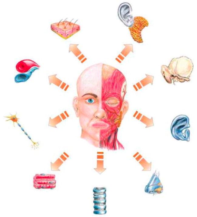
Engineering complex tissues requires a precisely formulated combination of cells, spatiotemporally released bioactive factors, and a specialized scaffold support system. Injectable materials, particularly those delivered in aqueous solution, are considered ideal delivery vehicles for cells and bioactive factors and can also be delivered through minimally invasive methods and fill complex 3D shapes. In this review, we examine injectable materials that form scaffolds or networks capable of both replacing tissue function early after delivery and supporting tissue regeneration over a time period of weeks to months. The use of these materials for tissue engineering within the craniofacial complex is challenging but ideal as many highly specialized and functional tissues reside within a small volume in the craniofacial structures and the need for minimally invasive interventions is desirable due to aesthetic considerations. Current biomaterials and strategies used to treat craniofacial defects are examined, followed by a review of craniofacial tissue engineering, and finally an examination of current technologies used for injectable scaffold development and drug and cell delivery using these materials.

Keywords: biomedical materials; drug delivery; hydrogels; polymeric materials; tissue engineering.


Figures

Figure 1
Figure 1
Craniofacial tissues needed in reconstruction. Reproduced with permission from [88], Springer Science+Business Media: European Archives of Otorhinolaryngology, Tissue Engineering in head and neck reconstructive surgery: what type of tissue do we need?, 264, 2007, 1344, Ulrich Reinhart Goessler, Jens Stern-Straeter, Katrin Riedel, Gregor M. Bran, Karl Hörmann, Frank Riedel, Figure 1.
Figure 2
Figure 2
These images show Silastic® implant fragments surgically retrieved from patients. The primary reason for retrieval was patient pain, likely secondary to implant failure. A and B show fracture lines within the implant, while C shows fraying and exposure of Dacron fibers that are intended to reinforce the implant. Reproduced with permission from [132], Journal of Oral and Maxillfacial Surgery, 66, J. N. A. R. Ferreira, C.-C. Ko, S. Myers, J. Swift, J. R. Fricton, Evaluation of Surgically Retrieved Temporomandibular Joint Alloplastic Implants: Pilot Study, 1112–1124, Copyright (2008), with permission from Elsevier
Figure 3
Figure 3
Cross sectional images of nerves regenerated in self assembling fibrin tubes. A 4 mm segmental defect was created in the dorsal root nerve of a rat and then bridged with a polymer tube implant. The gap was either left empty (A, B), filled with unmodified fibrin (C, D), or filled with fibrin modified with four peptides from the laminin family of adhesion molecules (E, F). The homogeneity of the nerve and alignment of the neuritis can be appreciated in the samples receiving peptide-modified fibrin bridges (A, C, E bar= 50 μm, B, D, F bar = 25 μm). Reprinted by permission from Macmillan Publishers Ltd: Nature Biotechnology (213), copyright (2000).
Figure 4
Figure 4
Focal adhesion formation of cells expressing green fluorescent protein labeled integrin (A–F) or yellow fluorescent protein labeled signal transduction protein (paxillin, G–L) in response to peptide amphiphiles expressing RGDS sequence(s). (A, G) branched peptide amphiphile with one cyclic RGDS, (B, H) branched peptide amphiphile with two RGDS, (C, I) branched peptide amphiphile with one RGDS, (D, J) branched peptide amphiphile with one d-RGDS, (E, K) linear peptide amphiphile with one RGDS, and (F, L) show cells on linear peptide amphiphile with one RGDS. Focal adhesions are seen as brightly fluorescent spots and demonstrate the presence of cellular organization during adhesion and migration in response to the presence of RGDS. This represents an example of how at the cellular level behavior can be regulated via substrate modification, a potentially powerful tool for regenerating of complex tissues. Reprinted from [216], Biomaterials, 28, H. Storrie, M. O. Guler, S. N. Abu-Amara, T. Volberg, M. Rao, B. Geiger, S. I. Stupp, Supramolecular crafting of cell adhesion, 4608-18, Copyright (2007), with permission from Elsevier.
Figure 5
Figure 5
Schematic representation of avidin induced self assembly of biotinylated PEG-PLA microparticles. Reproduced from [226].
Figure 6
Figure 6
Osteochondral tissue repair in rabbit knees 14 weeks after implantation with a bilayered scaffold (A, D, G – TGF-β1 releasing porous chondral and porous subchondral layers; B, E, H - TGF-β1 releasing porous chondral and nonporous subchondral layers; C, F, I porous chondral layer and nonporous subchondral layer). The boxed regions in (A–C) (2.5× magnification) are shown at higher magnifications in (D–F) (10×), and (G–I) (20×). Arrows point towards the joint surface, while columnar arrangements of chondrocytes, cell clusters, and cartilage fissures are respectively indicated by CO, CL, and FI. The images demonstrate the ability of a bilayered scaffold along with regionally specific growth factor release to regenerate multiple tissues within the same construct. Reproduced from [264].
Figure 7
Figure 7
Microcomputed tomography images of rat cranial bone defects treated with angiogenic, osteogenic or both growth factors. Figures A-D represent an untreated control group, angiogenic VEGF-treated group, osteogenic BMP-2-treated group, and a dual delivery group at 4 weeks. Blood vessels and bone are visible. Figures E–H represent control, VEGF, BMP-2 and dual groups at 12 weeks. Blood vessel formation was not evaluated at this time point. Bar represents 200 μm. Reprinted from [99], Bone, 43, Z S. Patel, S. Young, Y. Tabata, J.A. Jansen, M. Wong, A.G. Mikos, Dual delivery of an angiogenic and an osteogenic growth factor for bone regeneration in a critical size defect model, 931-40, Copyright (2008), with permission from Elsevier
Figure 8
Figure 8
Safranin O/fast green stained (left) and chondroitin-4-sulfate immunohistochemically stained cultures of human umbilical cord matrix (HUCM) derived stem cells and TMJ cartilage cells. HUCM were cultured in control (top left) and chondrogenically supplemented (top right) media for 4 weeks following initial culture in chondrogenic media. HUMCs produced more glycosaminoglycans as shown by both staining modalities than already differentiated TMJ chondrocytes, indicating the utility of stem cells in craniofacial tissue engineering compared to differentiated cells. Reproduced with permission from [283]. Copyright (2007) Mary Ann Liebert.
Figure 9
Figure 9
Cumulative secretion of osteopontin, a marker of osteogenic differentiation, from OPF hydrogel matrices with encapsulated MSCs. (A) MSCs encapsulated in OPF formulated from PEG with MW 10,000 Da and 3,000 Da were cultured in dexamethasone containing (+) and non-supplemented (−) culture media for 28 days. (B) Two types of OPF/MSC formulations maintained for 28 days in culture media without dexamethasone. The samples with higher molecular weight PEG (10K) underwent greater swelling than the less hydrophilic samples (PEG MW 3K), which led to enhanced osteogenic differentiation of encapsulated MSCs. Reproduced from [285].

References

    1. Axhausen W. J Bone Joint Surg Am. 1956;38-A:593. - PubMed
    1. Gutta R, Waite PD. The British journal of oral & maxillofacial surgery. 2007. - PubMed
    1. Louis PJ, Gutta R, Said-Al-Naief N, Bartolucci AA. J Oral Maxillofac Surg. 2008;66:235. - PubMed
    1. Sen MK, Miclau T. Injury. 200738(Suppl 1):S75. - PubMed
    1. Smolka W, Iizuka T. J Craniomaxillofac Surg. 2005;33:1. - PubMed

LinkOut - more resources