. 2009 Nov;39(11):3181-94.
doi: 10.1002/eji.200939496.

Evidence that cathelicidin peptide LL-37 may act as a functional ligand for CXCR2 on human neutrophils

Affiliations

Evidence that cathelicidin peptide LL-37 may act as a functional ligand for CXCR2 on human neutrophils

Zhifang Zhang et al. Eur J Immunol. 2009 Nov.

Abstract

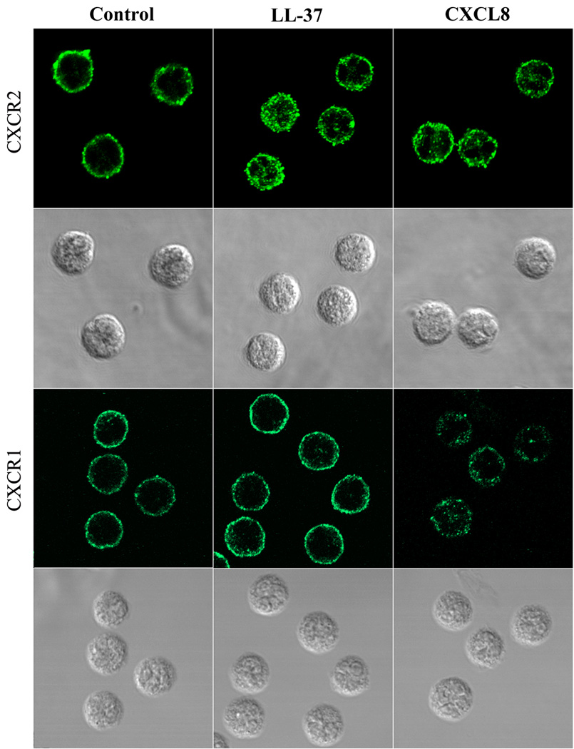
LL-37, derived from human cathelicidin, stimulates immune responses in neutrophils. Although FPR2 and P2X7 were proposed as LL-37 receptors, we have shown that among 21 neutrophil receptors only CXCR2 was down-regulated by LL-37. LL-37 functions similarly to CXCR2-specific chemokines CXCL1 and CXCL7 in terms of receptor down-regulation and intracellular calcium mobilization on freshly isolated neutrophils. Neutrophils pretreated with CXCL8, a chemokine that binds both CXCR1/2, completely blocked the calcium mobilization in response to LL-37, while LL-37 also partially inhibited (125)I-CXCL8 binding to neutrophils. SB225002, a selective CXCR2 antagonist, blocked LL-37-induced calcium mobilization and migration of neutrophils. LL-37 stimulates calcium mobilization in CXCR2-transfected HEK293 cells, CXCR2(+) THP-1 cells and monocytes, but not in CXCR1-transfected HEK293 cells. WKYMVm peptide (ligand for FPR2) does not block LL-37-stimulated calcium flux in either THP-1 (FPR2(-)) or monocytes (FPR2(high)), further confirming the specificity of LL-37 for CXCR2 and not FPR2. Among all ligands tested (ATP, BzATP, WKYMVm, CXCL1, and LL-37), only LL-37 stimulated migration of monocytes (CXCR2(+) and FPR2(+)) and migration was inhibited by the CXCR2 inhibitor SB225002. Moreover, CXCR2 but not CXCR1 was internalized in LL-37-treated neutrophils. Thus, our data provide evidence that LL-37 may act as a functional ligand for CXCR2 on human neutrophils.


Conflict of interest statement

Conflict of Interest: The authors declare no financial or commercial conflict of interest.

Figures

Figure 1
Figure 1. LL-37 down-regulates surface CXCR2 expression of human neutrophils
5×10⁶/mL neutrophils were incubated with LL-37 at four doses (1 µM, 2.5 µM, 5 µM, and 10 µM) for 3h or at 5 µM dose for four time points (0.5h, 1h, 2h, and 3h). The cells were washed and stained with anti-CXCR1 and anti-CXCR2 specific antibodies, analyzed by flow cytomety (GMFI, geometric mean of fluorescence intensity). (A) Dose response in 3 h stimulation. Time course of CXCR2 (B) and CXCR1 (C) expression in response to LL-37 (5 µM). Data show mean ± SE from four independent experiments. *p<0.05, **p<0.01, ***p<0.001 in comparison with CXCR2 expression of untreated control (unpaired Student’s t-test).
Figure 2
Figure 2. The patterns of surface CXCR1/2 expression of neutrophils treated with fMLF, WKYMVm, ATP, CXCL1, CXCL7, and CXCL8
5×10⁶/mL neutrophils were incubated with fMLF (A), FPR2 agonist WKYMVm (B), ATP (C), LL-37 (D), CXCL1 (E), CXCL7 (F), or CXCL8 (G) for 3h. The cells were washed and stained with anti-CXCR1-APC and anti-CXCR2-FITC (A–C and E–F) or anti-P2X7-FITC (D), and analyzed by flow cytomy (GMFI, geometric mean of fluorescence intensity). Data show mean ± SE from four independent experiments. * p<0.05 in comparison with control (D), All except (D), *p<0.05, **p<0.01, ***p<0.001 in comparison with CXCR2 expression of untreated control. #p<0.05, ##p<0.01, ###p<0.001 in comparison with CXCR1 expression of untreated control (unpaired Student’s t-test).
Figure 3
Figure 3. Effect of LL-37 on calcium mobilization of neutrophils
A–B. Neutrophils loaded with the calcium indicator indo-1 AM were suspended in PBS with calcium and magnesium, then treated with LL-37 (red line 1 µM, blue line 2.5 µM, green line 5 µM, and orange line 10 µM), CXCL1 (10 nM), CXCL7 (10 nM), CXCL8 (10 nM), ATP (5 µM), fMLF (100 nM) or WKYMVm (5 µM). C. Neutrophils loaded with the calcium indicator indo-1 AM were suspended in PBS without calcium and magnesium, then treated with LL37 (5 µM), WKYMVm (5 µM), or ATP (5 µM). The ratio of 405:485 nm was detected as intracellular calcium mobilization. The data shown are representative of four independent experiments. Arrows indicate time point at which reagents were applied to the cells.
Figure 4
Figure 4. Cross-desensitization of neutrophils by CXCL8 and LL-37
Neutrophils loaded with indo-1 AM were exposed to CXCL8 (10 nM). Cells were rechallenged 160 s later with a second stimulation by (A) LL-37 (5 µM), (B) CXCL7 (10 nM), (C) CXCL1 (10 nM), or (D) fMLF (100 nM). The ratio of 405:485 nm was detected as intracellular calcium mobilization. The data shown are representative of four independent experiments. Arrows indicate time point at which reagents were applied to the cells.
Figure 5
Figure 5. Competition of unlabelled LL-37, CXCL1, and CXCL8 with 125I-CXCL8 for binding to neutrophils
A. Specific binding of 125I-CXCL8 to receptors on human neutrophils. Neutrophils were incubated with increasing concentrations of 125I-CXCL8. Unspecific binding was determined in the presence of an excess (400-fold molar at 40 nM CXCL8). Specific binding was calculated by subtraction of unspecific binding from total binding. B. Competition of unlabeled LL-37, CXCL1, or CXCL8 with 125I-CXCL8 for binding to neutrophils. Neutrophils were incubated with 1.25 nM 125I-CXCL8 in the presence or in the absence of increasing concentrations of unlabeled LL-37, CXCL1 or CXCL8. Data were corrected for unspecific binding, determined in the presence of a 400-fold molar excess of the unlabeled CXCL8. Values were expressed as percentage of specific binding obtained with the labeled 125I-CXCL8 from the triplicate experiments. Data show mean ± SE of triplicate experiments.
Figure 6
Figure 6. SB225002 blocks LL-37-induced neutrophil calcium mobilization and migration
A–G. Neutrophils were loaded with indo-1 AM and exposed to SB225002 (10 µM), a selective CXCR2 antagonist. Cells were rechallenged 180 s later with a second stimulation by A. LL-37 (5 µM), B. CXCL7 (10 nM), C. CXCL1 (10 nM), D. CXCL8 (10 nM), E. fMLF (100 nM), F. ATP (5 µM), or G. WKYMVm (5 µM). The ratio of 405:485 nm was detected as intracellular calcium mobilization. The data shown are representative of four independent experiments with similar results. Arrows indicate time point at which reagents were applied to the cells. H. SB225002 blocks LL-37-induced neutrophil migration. Chemotaxis was measured in 24-well transwell system as described in the Materials and methods. Data show mean ± SE from three independent experiments performed in quadruplicate. *p<0.05, in comparison with untreated control; # p<0.05 in comparison with 5 µM LL-37 treatment (unpaired Student’s t-test).
Figure 7
Figure 7. Effect of LL-37 on calcium mobilization of HEK293/CXCR2/1 cells, THP-1 cells, and monocytes, and migration of THP-1 cells and monocytes
A–B. HEK293/CXCR1/2 cells loaded with the calcium indicator indo-1 AM were suspended in PBS with calcium and magnesium, then treated with CXCL8, LL-37, or CXCL1. C–D. THP-1 cells (C) or monocytes (D) were loaded with indo-1 AM and exposed to the first stimulus, then rechallenged with a second stimulus. The ratio of 405:485 nm was detected as intracellular calcium mobilization. The data shown are representative of three independent experiments. Arrows indicate the time point at which reagents were applied to the cells. E–F. THP-1 (E) and monocytes (F) show difference in response to LL-37 in chemotaxis. Chemotaxis was measured in 24-well transwell system as described in the Materials and methods. Ctrl (control), CXCL1 (50 nM), LL-37 (5 µM), WK (WKYMVm 5 µM), ATP (50 µM), BzATP (50 µM). Data show mean ± SE from three independent experiments performed in quadruplicate. ***p<0.001, in comparison with untreated control; ##p<0.01 in comparison with 5µM LL-37 treatment (unpaired Student’s t-test).
Figure 8
Figure 8. Confocal analysis of CXCR2 internalization in the LL-37 treated neutrophils
Neutrophils were incubated in the presence or absence of LL-37 (5 µM) or CXCL8 (50 nM) for 1 h. The cells were incubated with mouse anti-CXCR2 or anti-CXCR1 mAb, followed by staining with Alexa 488-conjugated rabbit anti-mouse IgG. Results are representative of three independent experiments.

References

    1. Sorensen O, Arnljots K, Cowland JB, Bainton DF, Borregaard N. The human antibacterial cathelicidin, hCAP-18, is synthesized in myelocytes and metamyelocytes and localized to specific granules in neutrophils. Blood. 1997;90:2796–2803. - PubMed
    1. Frohm Nilsson M, Sandstedt B, Sorensen O, Weber G, Borregaard N, Stahle-Backdahl M. The human cationic antimicrobial protein (hCAP18), a peptide antibiotic, is widely expressed in human squamous epithelia and colocalizes with interleukin-6. Infect Immun. 1999;67:2561–2566. - PMC - PubMed
    1. Agerberth B, Charo J, Werr J, Olsson B, Idali F, Lindbom L, Kiessling R, Jornvall H, Wigzell H, Gudmundsson GH. The human antimicrobial and chemotactic peptides LL-37 and alpha-defensins are expressed by specific lymphocyte and monocyte populations. Blood. 2000;96:3086–3093. - PubMed
    1. Sorensen OE, Follin P, Johnsen AH, Calafat J, Tjabringa GS, Hiemstra PS, Borregaard N. Human cathelicidin, hCAP-18, is processed to the antimicrobial peptide LL-37 by extracellular cleavage with proteinase 3. Blood. 2001;97:3951–3959. - PubMed
    1. Larrick JW, Hirata M, Balint RF, Lee J, Zhong J, Wright SC. Human CAP18: a novel antimicrobial lipopolysaccharide-binding protein. Infect Immun. 1995;63:1291–1297. - PMC - PubMed

Publication types

LinkOut - more resources