Review
. 2009 Sep;87(3):222-31.
doi: 10.1002/bdrc.20155.

Lymphatic development

Affiliations
Review

Lymphatic development

Matthew G Butler et al. Birth Defects Res C Embryo Today. 2009 Sep.

Abstract

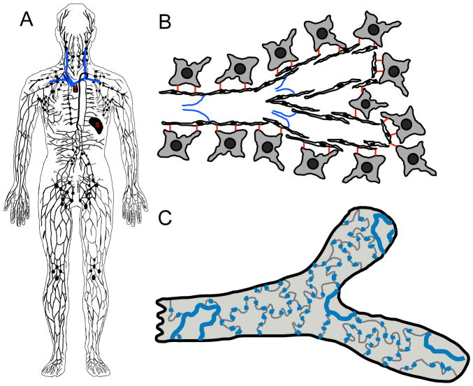
The lymphatic system is essential for fluid homeostasis, immune responses, and fat absorption, and is involved in many pathological processes, including tumor metastasis and lymphedema. Despite its importance, progress in understanding the origins and early development of this system has been hampered by lack of defining molecular markers and difficulties in observing lymphatic cells in vivo and performing genetic and experimental manipulation of the lymphatic system. Recent identification of new molecular markers, new genes with important functional roles in lymphatic development, and new experimental models for studying lymphangiogenesis has begun to yield important insights into the emergence and assembly of this important tissue. This review focuses on the mechanisms regulating development of the lymphatic vasculature during embryogenesis.


Figures

Figure 1
Figure 1
Characteristics of the lymphatic vasculature. (A) An overview of the human lymphatic system, including lymphatic vessels, lymph nodes, and lymphoid tissue (s-spleen, t-thymus). Major veins into which the lymphatics drain are shown in blue. (B) The lymphatic endothelial cells attach directly to the extracellular matrix and surrounding cells via anchoring filaments (red). Valves (blue) prevent lymph reflux to promote unidirectional lymph propulsion. Note the extensive overlap of adjacent endothelial cells in lymphatic capillaries. (C) Surface view of a lymphatic capillary emphasizing the loose, button-like intercellular junctions (blue).
Figure 2
Figure 2
Anatomical and functional characterization of zebrafish lymphatic vessels. (A, B) Diagram of the superficial (A) and major conserved superficial (blue) and deeper (green) lymphatics (B) of the salamander, including lateral (L) and vertebral (Vt) superficial, and spinal (S) and collateral cardinal (C) deeper lymphatics. (C) 5 week old Tg(fli1:EGFP)yl zebrafish (green vessels) with fluorescent microspheres injected into the lymphatic vascular system (red lymphatics), showing major trunk superficial (small arrows) and deeper (large arrows) lymphatics similar to those in panel A. (D, E) Berlin blue dye injected into the lymphatic vascular system of a 5 week zebrafish (D) with explanatory diagram (E). As in other vertebrates, longitudinal lateral lymphatic (LL), pectoral lymphatic (PL), and facial lymphatic (FL) vessels come together to drain into the common cardinal vein (CCV). (F–H) Confocal imaging of an 18 dpf Tg(fli1:EGFP)yl zebrafish (green) injected subcutaneously with 2 Md rhodamine-dextran (red). (F) Subcutaneously injected rhodamine-dextran is taken up by lymphatic vessels and drains into the thoracic duct (small arrow), but does not label the adjacent dorsal aorta (large arrow). (G) Numerous rhodamine-dextran labeled vessels (red) are visible between the blood vessels (green). (H) Higher magnification image of blind-ended (arrows) rhodamine-dextran labeled vessels. Scale bars = 100 μm (C), 20 μm (F), 50 μm (G), or 100 μm (H). Images from Yaniv et al., Nature Medicine 12,711–716 (2006).
Figure 3
Figure 3
Assembly and origins of the zebrafish lymphatic thoracic duct visualized directly in the zebrafish. (A, B) Two-photon time-lapse imaging of formation of the trunk thoracic duct, collected from 2–4 dpf Tg(fli1:EGFP)yl zebrafish. Diagrams of the areas imaged (red box) at time zero are shown at top. Selected frames from the time-lapse sequences are shown below, with the lymphatic sprouts highlighted in red. (A) A lymphatic sprout for the thoracic duct emerging and then growing rostrally and caudally ventral to the dorsal aorta. (B) Lymphatic vessel sprouts growing across the trunk and merging ventral to the dorsal aorta. (C–E) Two-photon time-lapse imaging of EGFP-positive endothelial cell nuclei migrating to and incorporating into the thoracic duct in the trunk of a 2–4 dpf Tg(fli1:nEGFP)y7 zebrafish. (C) Explanatory diagram showing that two cells (yellow and red) migrate ventrally from the parachordal vessel to contribute to the lymphatic thoracic duct. The diagram shows the location of the cells and their daughters at different time points (in hours). (D, E) Actual images from the time-lapse sequence showing the positions of lymphatic progenitor cells (1, 2) and their daughters (1a, 1b, 2a, 2b) at time zero (D) and 30.5 hr after the start of the time lapse sequence (E). Scale bars = 25 μm (A, B), 50 μm (D, E). Images from Yaniv et al., Nature Medicine 12,711–716 (2006).
Figure 4
Figure 4
Simplified speculative upstream and downstream Prox1 (A) and Vegfr3 (B) signaling pathways in lymphatic endothelium. Solid arrows show activation and perpendicular lines show inhibition. Solid arrows show direct interactions, whereas dashed arrows show undefined interactions. The pathways shown are not comprehensive, nor are their functional outputs as discrete as presented (e.g., VegfC promotes both survival and migration of Vegfr3 expressing LRC).

References

    1. Abtahian F, Guerriero A, Sebzda E, Lu MM, Zhou R, Mocsai A, Myers EE, Huang B, Jackson DG, Ferrari VA, Tybulewicz V, Lowell CA, Lepore JJ, Koretzky GA, Kahn ML. Regulation of blood and lymphatic vascular separation by signaling proteins SLP-76 and Syk. Science (New York, NY. 2003;299(5604):247–251. - PMC - PubMed
    1. Achen MG, Jeltsch M, Kukk E, Makinen T, Vitali A, Wilks AF, Alitalo K, Stacker SA. Vascular endothelial growth factor D (VEGF-D) is a ligand for the tyrosine kinases VEGF receptor 2 (Flk1) and VEGF receptor 3 (Flt4) Proceedings of the National Academy of Sciences of the United States of America. 1998;95(2):548–553. - PMC - PubMed
    1. Aselli G. De Lacteibus sive Lacteis Venis, Quarto Vasorum Mesarai corum Genere novo invento. Milan: Mediolani; 1627.
    1. Baldwin ME, Halford MM, Roufail S, Williams RA, Hibbs ML, Grail D, Kubo H, Stacker SA, Achen MG. Vascular endothelial growth factor D is dispensable for development of the lymphatic system. Molecular and cellular biology. 2005;25(6):2441–2449. - PMC - PubMed
    1. Baluk P, Fuxe J, Hashizume H, Romano T, Lashnits E, Butz S, Vestweber D, Corada M, Molendini C, Dejana E, McDonald DM. Functionally specialized junctions between endothelial cells of lymphatic vessels. The Journal of experimental medicine. 2007;204(10):2349–2362. - PMC - PubMed

LinkOut - more resources