Review
. 2009 Nov;19(6):359-78.
doi: 10.1002/rmv.630.

Viruses and autophagy

Affiliations
Review

Viruses and autophagy

Sagar B Kudchodkar et al. Rev Med Virol. 2009 Nov.

Abstract

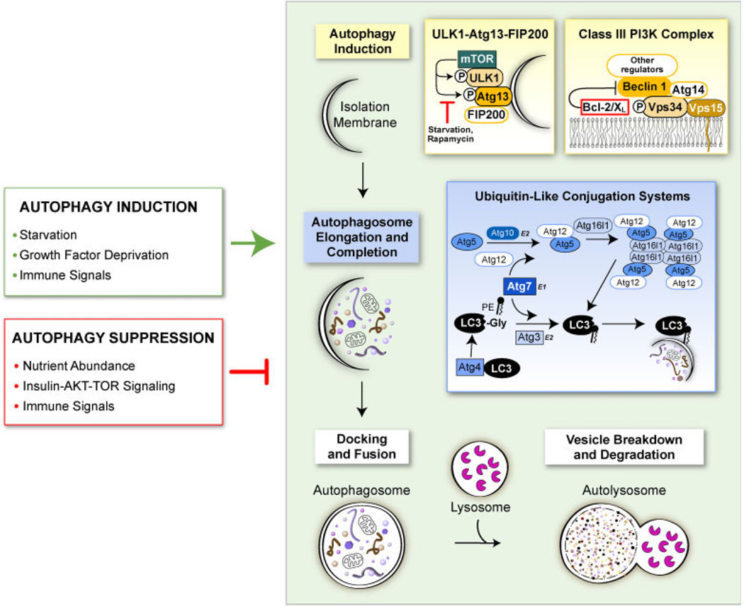
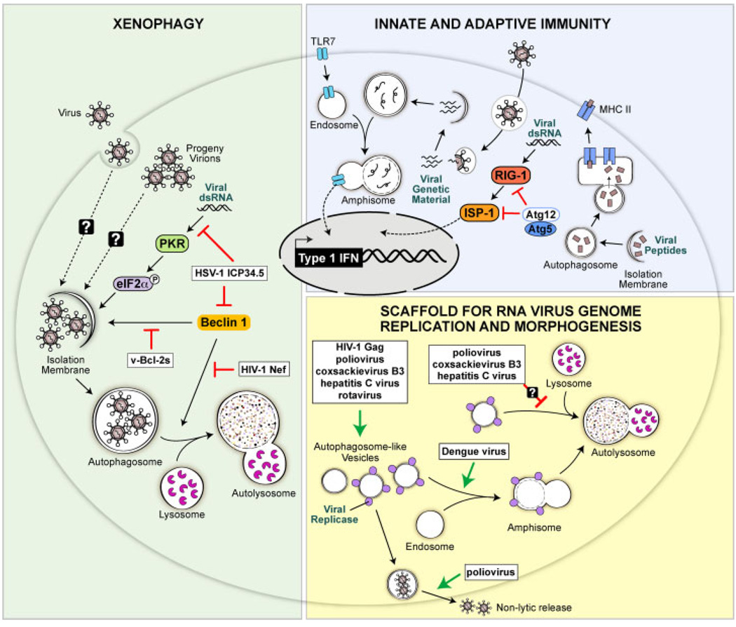
Autophagy is an evolutionarily conserved intracellular process by which bulk cytoplasm is enveloped inside a double-membraned vesicle and shuttled to lysosomes for degradation. Within the last 15 years, the genes necessary for the execution of autophagy have been identified and the number of tools for studying this process has grown. Autophagy is essential for tissue homeostasis and development and defective autophagy is associated with a number of diseases. As intracellular parasites, during the course of an infection, viruses encounter autophagy and interact with the proteins that execute this process. Autophagy and/or autophagy genes likely play both anti-viral and pro-viral roles in the life cycles and pathogenesis of many different virus families. With respect to anti-viral roles, the autophagy proteins function in targeting viral components or virions for lysosomal degradation in a process termed xenophagy, and they also play a role in the initiation of innate and adaptive immune system responses to viral infections. Consistent with this anti-viral role of host autophagy, some viruses encode virulence factors that interact with the host autophagy machinery and block the execution of autophagy. In contrast, other viruses appear to utilise components of the autophagic machinery to foster their own intracellular growth or non-lytic cellular egress. As the details of the role (s) of autophagy in viral pathogenesis become clearer, new anti-viral therapies could be developed to inhibit the beneficial and enhance the destructive aspects of autophagy on the viral life cycle.


Figures

Figure 1
Figure 1
Scheme of the autophagy pathway. The process of autophagy begins with the nucleation of an autophagic isolation membrane that eventually encloses the cytoplasmic and/or microbial cargo destined for lysosomal degradation. The mammalian target of rapamycin (mTOR) kinase plays a major role in suppressing autophagy induction by binding to ULK1-Atg13-FIP200 complexes and hyper-phosphorylating ULK1 and Atg13. Under autophagy promoting conditions such as starvation, mTOR does not associate with the ULK1-Atg13-FIP200 complex, leaving ULK1 and Atg13 hypo-phosphorylated, which allows the isolation membrane to expand. Vesicle nucleation is also promoted through the activation of the class III phosphatidylinositol 3-phosphate (PIP3) kinase Vps34 which contains the essential mammalian autophagy proteins Beclin 1 and Atg14. The Vps34-Beclin 1-Atg14 complex creates PIP3 residues on the isolation membrane that serve as docking sites for other autophagy-promoting proteins. Other proteins bind to this minimal class III PI3K complex to regulate autophagy and other membrane-trafficking processes, including Bcl-2 family members that function as important inhibitors of autophagy. The second major step of autophagy involves the elongation of the isolation membrane to form an autophagosome which has two phospholipid bilayer membranes with the selected cargo sequestered within the inner membrane. Two ubiquitin-like conjugation systems mediate this step by modifying two ubiquitin-like molecules, Atg5 and LC3, so that they can associate with the isolation membrane and promote its curvature and expansion. The last steps of autophagy involve the docking of completed autophagosomes with lysosomes and the lysosomal enzyme-mediated breakdown and degradation of the inner membrane of the autophagosome and its constituents. Autophagy is triggered and inhibited in cells in response to a diverse set of stimuli including nutrient availability, growth factor mediated-signaling and immune signals generated in response to microbes (green and red boxes)
Figure 2
Figure 2
Anti-viral and pro-viral functions of autophagy. (Green-shaded region) Autophagy may function as a cell-intrinsic anti-viral mechanism by directly capturing virions and/or viral components and targeting them for degradation in lysosomes, a process known as xenophagy. The PKR-eIF2α signaling pathway and the autophagy protein Beclin 1 function in promoting xenophagy, and some viruses have evolved to encode gene products to block the autophagy-promoting activities of these proteins. The HSV-1 ICP34.5 protein blocks both PKR- and Beclin 1-mediated activation of autophagy and viral Bcl-2 homologues found in γ-herpesviruses block Beclin 1-mediated autophagy. The Nef protein of HIV-1 blocks the ability of Beclin 1 to promote autophagosome maturation into autolysosomes. (Blue-shaded region) Autophagy and autophagy genes also exert anti-viral functions by collaborating with the innate and adaptive immune systems. Autophagy delivers viral genetic material from SV and vesicular stomatits virus to endosomes where the ssRNA-sensing toll like receptor 7 resides thus initiating anti-viral Type I interferon signaling. Autophagosomes also target viral peptides to MHC class II loading compartments, thereby enhancing CD4 T cell responses to these antigens. In contrast to promoting innate immunity, the autophagy proteins Atg5 and Atg12 may function to dampen innate immune responses to viruses by binding to RIG-I, a sensor of foreign genetic material, and blocking signaling downstream of RIG-I. (Yellow-shaded region) For some viruses, the autophagy machinery may function in a pro-viral capacity by creating double membrane structures that serve as scaffolds for the replication of viral genomes or as sites of virion morphogenesis. Several RNA viruses including poliovirus, coxsackievirus B3, hepatitis C virus and rotavirus have been shown to induce double-membraned, autophagosome like structures in infected cells, and replicases from these viruses associate with these structures. Poliovirus may also use these structures to promote the non-lytic release of new virions from cells. All of these viruses presumably have means to block the maturation of these structures into destructive autolysosomes. The Gag protein of HIV-1 also appears to be involved in forming early stage autophagosomes in macrophages which enhances Gag processing and HIV yields from macrophages through an unknown mechanism

References

    1. Levine B, Klionsky DJ. Development by self-digestion: molecular mechanisms and biological functions of autophagy. Dev Cell. 2004;6(4):463–477. - PubMed
    1. Levine B, Kroemer G. Autophagy in the pathogenesis of disease. Cell. 2008;132(1):27–42. - PMC - PubMed
    1. Xie Z, Klionsky DJ. Autophagosome formation: core machinery and adaptations. Nat Cell Biol. 2007;9(10):1102–1109. - PubMed
    1. Hosokawa N, Hara T, Kaizuka T, et al. Nutrient-dependent mTORC1 association with the ULK1-Atg13-FIP200 complex required for autophagy. Mol Biol Cell. 2009;20(7):1981–1991. - PMC - PubMed
    1. Talloczy Z, Jiang W, Virgin HW, 4th, et al. Regulation of starvation- and virus-induced autophagy by the eIF2a kinase signaling pathway. Proc Natl Acad Sci USA. 2002;99(1):190–195. - PMC - PubMed

Publication types