

Aptamers generated from cell-SELEX for molecular medicine: a chemical biology approach
- PMID: 19751057
- PMCID: PMC2808443
- DOI: 10.1021/ar900101s
Aptamers generated from cell-SELEX for molecular medicine: a chemical biology approach
Abstract
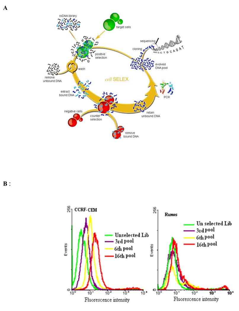
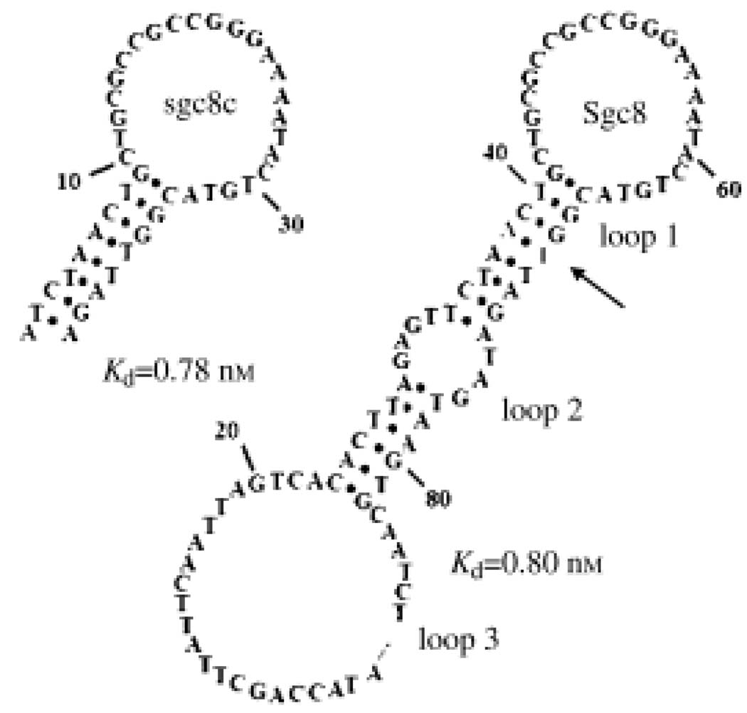
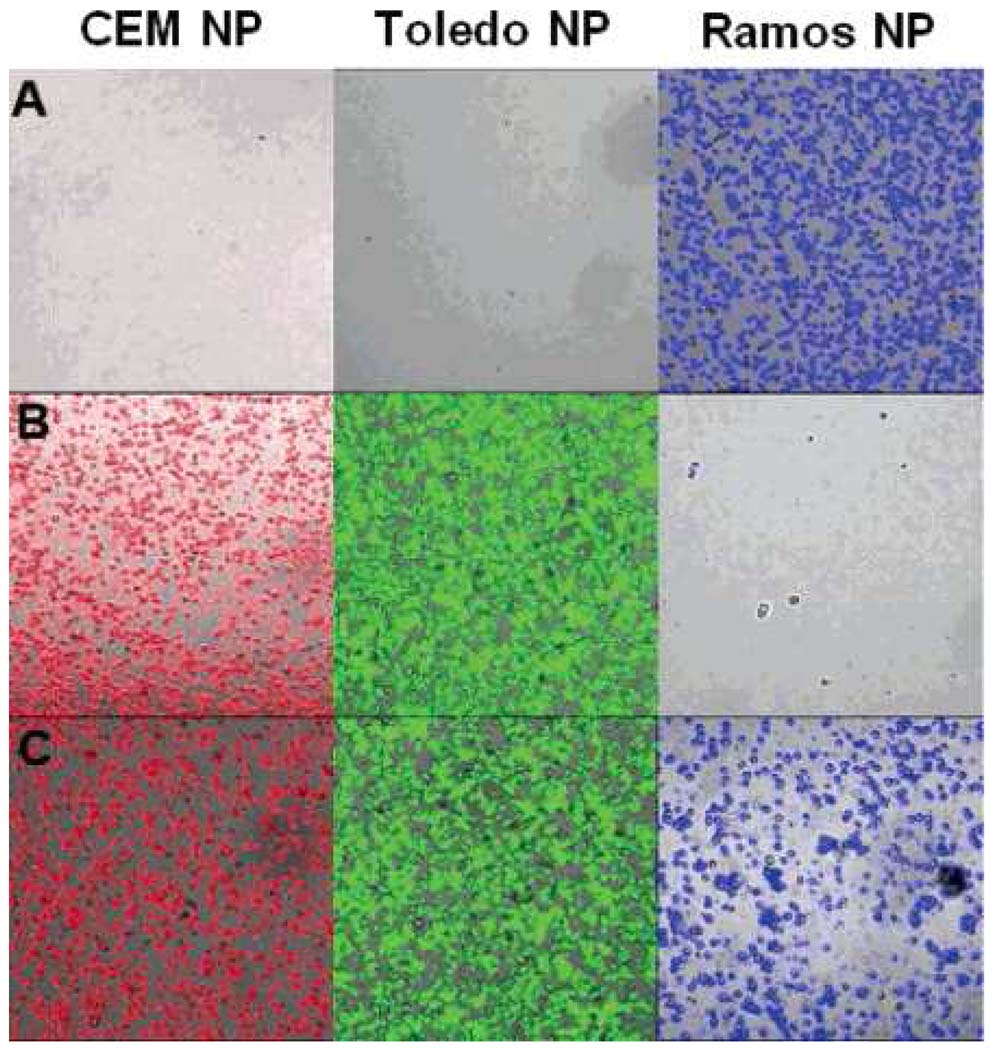
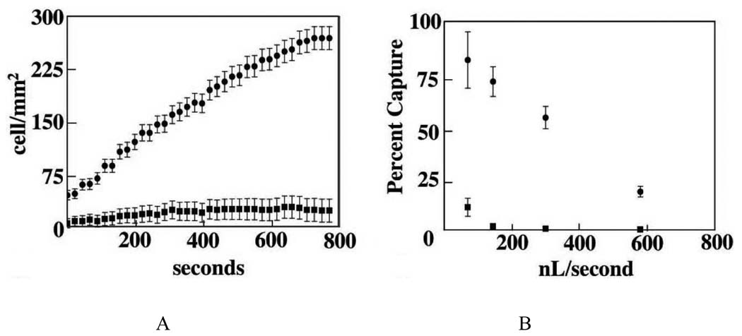
Molecular medicine is an emerging field focused on understanding the molecular basis of diseases and translating this information into strategies for diagnosis and therapy. This approach could lead to personalized medical treatments. Currently, our ability to understand human diseases at the molecular level is limited by the lack of molecular tools to identify and characterize the distinct molecular features of the disease state, especially for diseases such as cancer. Among the new tools being developed by researchers including chemists, engineers, and other scientists is a new class of nucleic acid probes called aptamers, which are ssDNA/RNA molecules selected to target a wide range of molecules and even cells. In this Account, we will focus on the use of aptamers, generated from cell-based selections, as a novel molecular tool for cancer research. Cancers originate from mutations of human genes. These genetic alterations result in molecular changes to diseased cells, which, in turn, lead to changes in cell morphology and physiology. For decades, clinicians have diagnosed cancers primarily based on the morphology of tumor cells or tissues. However, this method does not always give an accurate diagnosis and does not allow clinicians to effectively assess the complex molecular alterations that are predictive of cancer progression. As genomics and proteomics do not yet allow a full access to this molecular knowledge, aptamer probes represent one effective and practical avenue toward this goal. One special feature of aptamers is that we can isolate them by selection against cancer cells without prior knowledge of the number and arrangement of proteins on the cellular surface. These probes can identify molecular differences between normal and tumor cells and can discriminate among tumor cells of different classifications, at different disease stages, or from different patients. This Account summarizes our recent efforts to develop aptamers through cell-SELEX for the study of cancer and apply those aptamers in cancer diagnosis and therapy. We first discuss how we select aptamers against live cancer cells. We then describe uses of these aptamers. Aptamers can serve as agents for molecular profiling of specific cancer types. They can also be used to modify therapeutic reagents to develop targeted cancer therapies. Aptamers are also aiding the discovery of new cancer biomarkers through the recognition of membrane protein targets. Importantly, we demonstrate how molecular assemblies can integrate the properties of aptamers and, for example, nanoparticles or microfluidic devices, to improve cancer cell enrichment, detection and therapy.
Figures
References
-
- Tuerk C, Gold L. Systematic Evolution of Ligands by Exponential Enrichment: RNA Ligands to Bacteriophage T4 DNA Polymerase. Science. 1990;249:505–510. - PubMed
-
- Ellington AD, Szostak JW. In vitro Selection of RNA Molecules that Bind Specific Ligands. Nature. 1990;346:818–822. - PubMed
-
- Osborne SE, Matsumura I, Ellington AD. Aptamers as Therapeutic and Diagnostic Reagents: Problems and Prospects. Curr. Opin. Chem. Biol. 1997;1:5–9. - PubMed
-
- Famulok M, Hartig JS, Mayer G. Functional Aptamers and Aptazymes in Biotechnology, Diagnostics, and Therapy. Chem. Rev. 2007;107:3715–3743. - PubMed
-
- Navani NK, Li Y. Nucleic Acid Aptamers and Enzymes as Sensors. Curr. Opin. Chem. Biol. 2006;10:272–281. - PubMed
Publication types
MeSH terms
Substances
Grants and funding
LinkOut - more resources
Full Text Sources
Other Literature Sources
Research Materials
