. 2009 Sep 14:9:325.
doi: 10.1186/1471-2407-9-325.

Expression analysis of E-cadherin, Slug and GSK3beta in invasive ductal carcinoma of breast

Affiliations

Expression analysis of E-cadherin, Slug and GSK3beta in invasive ductal carcinoma of breast

Chandra P Prasad et al. BMC Cancer. .

Abstract

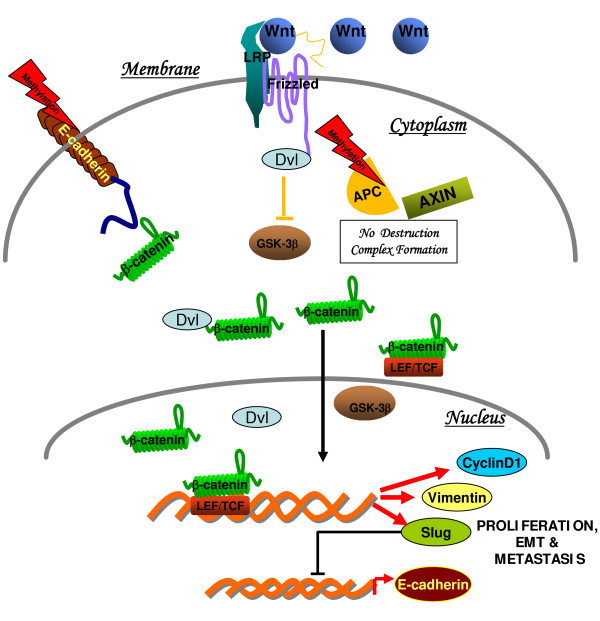
Background: Cancer progression is linked to a partially dedifferentiated epithelial cell phenotype. The signaling pathways Wnt, Hedgehog, TGF-beta and Notch have been implicated in experimental and developmental epithelial mesenchymal transition (EMT). Recent findings from our laboratory confirm that active Wnt/beta-catenin signaling is critically involved in invasive ductal carcinomas (IDCs) of breast.

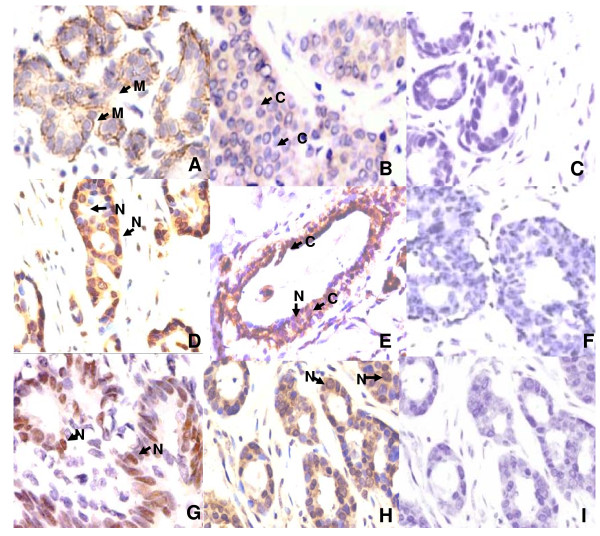
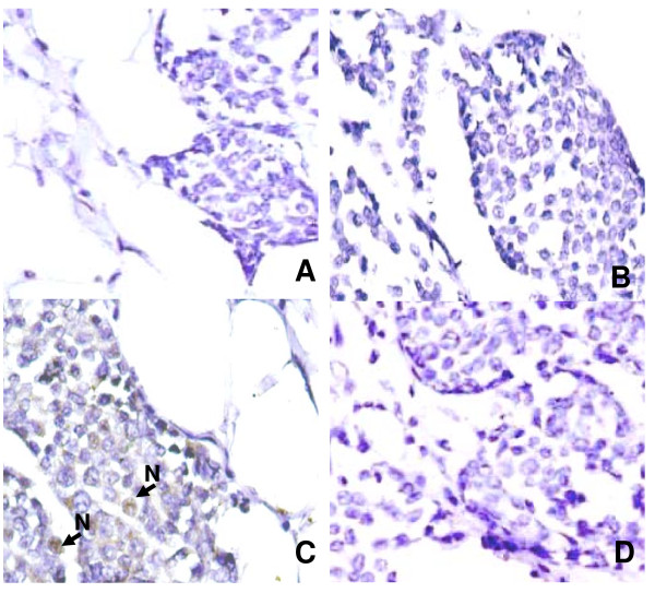
Methods: In the current study, we analyzed the expression patterns and relationships between the key Wnt/beta-catenin signaling components- E-cadherin, Slug and GSK3beta in IDCs of breast.

Results: Of the 98 IDCs analyzed, 53 (54%) showed loss/or reduced membranous staining of E-cadherin in tumor cells. Nuclear accumulation of Slug was observed in 33 (34%) IDCs examined. Loss or reduced level of cytoplasmic GSK3beta expression was observed in 52/98 (53%) cases; while 34/98 (35%) tumors showed nuclear accumulation of GSK3beta. Statistical analysis revealed associations of nuclear Slug expression with loss of membranous E-cadherin (p = 0.001); nuclear beta-catenin (p = 0.001), and cytoplasmic beta-catenin (p = 0.005), suggesting Slug mediated E-cadherin suppression via the activation of Wnt/beta-catenin signaling pathway in IDCs. Our study also demonstrated significant correlation between GSK3beta nuclear localization and tumor grade (p = 0.02), suggesting its association with tumor progression.

Conclusion: The present study for the first time provided the clinical evidence in support of Wnt/beta-catenin signaling upregulation in IDCs and key components of this pathway - E-cadherin, Slug and GSK3beta with beta-catenin in implementing EMT in these cells.


Figures

Figure 1
Figure 1
Schematic diagram showing Wnt/β-catenin signaling in Invasive ductal carcinomas of breast: On activation of Wnt signaling, disheveled (Dvl) prevents degradation of β-catenin, possibly through the recruitment of GBP/Frat-1, which in turn displaces GSK3β from the destruction complex. Adenomatous Polyposis Coli (APC), an important component of Wnt signaling was also found to downregulated by promoter methylation, as one of the mechanism [21]. Stabilized β-catenin enters the nucleus and associates with T cell factor (TCF)/lymphoid enhancer factor (LEF) transcription factors, which leads to the transcription of Wnt target genes such as cyclin D1, vimentin and slug [20,21]. Mechanisms attributed in the downregulation of E-cadherin are DNA methylation/or by transcriptional suppression via snail/or slug, thereby releasing membrane bound β-catenin into the cytosol [21].
Figure 2
Figure 2
Expression of E-cadherin, Slug and GSK3β proteins in invasive ductal carcinomas of breast. (A) Strong membranous E-cadherin expression in normal breast tissue (B) Loss of E-cadherin membranous localization in tumor cells. (C) Loss of Slug protein in normal breast (D) Nuclear accumulation of Slug in IDC (E) GSK3β expression in normal breast epithelium (F) IDC showing loss cytoplasmic GSK3β (G) GSK3β nuclear localization in tumor cells; and (H&I) Nuclear accumulation of Slug and loss of E-cadherin immunostaining examined on the adjacent sections of the same tumor. (A-I, original magnification × 400). Arrows show Membranous (M), Nuclear (N) and cytoplasmic (C) localization of the proteins.
Figure 3
Figure 3
Expression pattern of E-cadherin, Slug and GSK3β proteins in invasive lobular carcinomas of breast. (A& B) Invasive lobular carcinomas showing loss of E-cadherin staining (C) Invasive lobular carcinoma showing nuclear staining for Slug (D) ILC showing complete loss of GSK3β protein. (A-D, original magnification × 400).

References

    1. Report of the National Cancer Registry Programme. Ind Council Med Res New Delhi. 2001. p. 22.
    1. Mittra I. Overview of breast cancer in India. European J Cancer. 2006;4(Suppl):180.
    1. Shook D, Keller R. Mechanisms, mechanics and function of epithelial-mesenchymal transitions in early development. Mech Dev. 2003;120:1351–83. doi: 10.1016/j.mod.2003.06.005. - DOI - PubMed
    1. Wu SY, Ferkowicz M, McClay DR. Ingression of primary mesenchyme cells of the sea urchin embryo: a precisely timed epithelial mesenchymal transition. Birth Defects Res C Embryo Today. 2007;81:241–252. doi: 10.1002/bdrc.20113. - DOI - PubMed
    1. Neth P, Ries C, Karow M, Egea V, Ilmer M, Jochum M. The Wnt signal transduction pathway in stem cells and cancer cells: influence on cellular invasion. Stem Cell Rev. 2007;3:18–29. doi: 10.1007/s12015-007-0001-y. - DOI - PubMed

Publication types

MeSH terms