Review
. 2009 Sep-Oct;78(2-3):185-94.
doi: 10.1016/j.diff.2009.08.004.

A home away from home: challenges and opportunities in engineering in vitro muscle satellite cell niches

Affiliations
Review

A home away from home: challenges and opportunities in engineering in vitro muscle satellite cell niches

Benjamin D Cosgrove et al. Differentiation. 2009 Sep-Oct.

Abstract

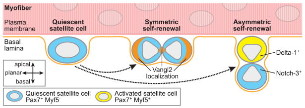
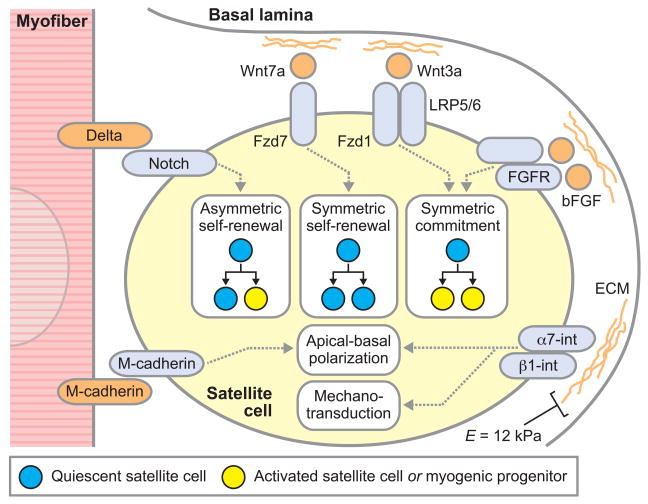
Satellite cells are skeletal muscle stem cells with a principal role in postnatal skeletal muscle regeneration. Satellite cells, like many tissue-specific adult stem cells, reside in a quiescent state in an instructive, anatomically defined niche. The satellite cell niche constitutes a distinct membrane-enclosed compartment within the muscle fiber, containing a diversity of biochemical and biophysical signals that influence satellite cell function. A major limitation to the study and clinical utility of satellite cells is that upon removal from the muscle fiber and plating in traditional plastic tissue culture platforms, their muscle stem cell properties are rapidly lost. Clearly, the maintenance of stem cell function is critically dependent on in vivo niche signals, highlighting the need to create novel in vitro microenvironments that allow for the maintenance and propagation of satellite cells while retaining their potential to function as muscle stem cells. Here, we discuss how emerging biomaterials technologies offer great promise for engineering in vitro microenvironments to meet these challenges. In engineered biomaterials, signaling molecules can be presented in a manner that more closely mimics cell-cell and cell-matrix interactions, and matrices can be fabricated with diverse rigidities that approximate in vivo tissues. The development of in vitro microenvironments in which niche features can be systematically modulated will be instrumental not only to future insights into muscle stem cell biology and therapeutic approaches to muscle diseases and muscle wasting with aging, but also will provide a paradigm for the analysis of numerous adult tissue-specific stem cells.


Figures

Figure 1
Figure 1. Modes of satellite cell self-renewal
Symmetric stem cell self-renewal leads to two daughter cells retaining stem cell function, and thus an expansion of the stem cell pool, which is necessary for tissue regeneration following injury or disease. In comparison, asymmetric stem cell self-renewal yields one daughter stem cell and one differentiated cell and therefore is sufficient to maintain the stem cell pool under homeostatic conditions (Morrison and Kimble, 2006). While satellite cells could, in theory, arise from reversion of a differentiated cell back to a stem cell phenotype (Zammit et al., 2004), this schematic illustrates only the modes of quiescent satellite cell self-renewal supported by recent experimental findings using Cre-loxP-mediated cell lineage tracing (Le Grand et al., 2009; Kuang et al., 2007). In Myf5-Cre/ROSA26-YFP transgenic mice, cells that have expressed Myf5, a marker of satellite cell activation which is expressed concomitant with a substantial reduction in potential to contribute to muscle regeneration, express the fluorescent protein YFP. In myofibers isolated from these transgenic mice, quiescent Pax7+Myf5 satellite cells can give rise to either (i) two Pax7+Myf5 quiescent satellite cells (symmetric self-renewal); (ii) one Pax7+Myf5 quiescent satellite cell and one Pax7+Myf5+ activated satellite cell (asymmetric self-renewal); or (iii) two Pax7+Myf5+ activated satellite cells (commitment, not shown in diagram). Moreover, daughter cells from satellite cell divisions are orientated relative to the myofiber plasma membrane and basal lamina in a manner consistent with their commitment status. In the case of symmetric self-renewal, the two daughter quiescent satellite cells are in planar alignment with the myofiber and have been shown to localize components of the planar cell polarity pathway, including Vangl2, in a parallel pattern. In the case of asymmetric self-renewal, the quiescent daughter cell remains in contact with the basal lamina (basal position) and the activated daughter cell is adjacent to the myofiber plasma membrane (apical position), positioned for possible fusion with the myofiber. The apical activated satellite cells express the Notch ligand Delta-1 and basal quiescent satellite cells express the receptor Notch-3, suggesting that the more differentiated progeny activate Notch signaling in adjacent quiescent satellite cells that can contribute to maintenance of their quiescent state. Emerging evidence suggests that these modes of self-renewal are controlled by specific molecular components of the satellite cell niche (see Figure 2).
Figure 2
Figure 2. Niche components that regulate satellite cell fate and function
Satellite cells reside in a niche between the myofiber plasma membrane and basal lamina consisting of a diversity of biochemical and biophysical signals that dictate their fate and function. This diagram illustrates some key niche components, their recognition by satellite cell receptors, and roles in regulating satellite cell self-renewal, proliferation, commitment, and polarity. Satellite cell niche components can be asymmetrically distributed, with factors associated with the myofiber plasma membrane presented to the apical surface of satellite cells and factors associated with the basal lamina presented to the basal surface of satellite cells. The myofiber basal lamina is a network of extracellular matrix components, including type IV collagen, laminin, fibronectin, and proteoglycans. These basal lamina ECM components facilitate satellite cell adhesion via integrin binding and activate integrin-related signaling to establish, in concert with myofiber-associated signals, apical-basal polarity in satellite cells and allow satellite cells to sense the mechanical stiffness of their microenvironment. (Resting healthy skeletal muscle has an elastic modulus (E) of ~12 kPa.) Whether apical-basal polarity or sensing of the microenvironmental mechanical stiffness provide critical determinants of satellite cell fate and function remain to be elucidated. Proteoglycan components of the ECM can bind an array of soluble glycoproteins, including bFGF and Wnt ligands, which are derived from systemic, satellite cell, or myofiber sources, localizing these factors to the satellite cell basal surface. Moreover, cell surface ligands, including the Delta family of ligands for the Notch receptor, are expressed by myofibers and satellite cells themselves and can stimulate satellite cells by engagement with their cognate receptors. Artificial satellite cell niches will allow a systematic analysis of the influences of these specific components on stem cell fate and function.

References

    1. Alberti K, Davey RE, Onishi K, George S, Salchert K, Seib FP, Bornhauser M, Pompe T, Nagy A, Werner C, Zandstra PW. Functional immobilization of signaling proteins enables control of stem cell fate. Nat Methods. 2008;5:645–650. - PubMed
    1. Alexakis C, Partridge T, Bou-Gharios G. Implication of the satellite cell in dystrophic muscle fibrosis: a self-perpetuating mechanism of collagen overproduction. Am J Physiol Cell Physiol. 2007;293:C661–669. - PubMed
    1. Beauchamp JR, Heslop L, Yu DS, Tajbakhsh S, Kelly RG, Wernig A, Buckingham ME, Partridge TA, Zammit PS. Expression of CD34 and Myf5 defines the majority of quiescent adult skeletal muscle satellite cells. J Cell Biol. 2000;151:1221–1234. - PMC - PubMed
    1. Beauchamp JR, Morgan JE, Pagel CN, Partridge TA. Dynamics of myoblast transplantation reveal a discrete minority of precursors with stem cell-like properties as the myogenic source. J Cell Biol. 1999;144:1113–1122. - PMC - PubMed
    1. Benchaouir R, Meregalli M, Farini A, D’Antona G, Belicchi M, Goyenvalle A, Battistelli M, Bresolin N, Bottinelli R, Garcia L, Torrente Y. Restoration of human dystrophin following transplantation of exon-skipping-engineered DMD patient stem cells into dystrophic mice. Cell Stem Cell. 2007;1:646–657. - PubMed

Publication types