Comparative Study
. 2009 Dec;77(12):5478-85.
doi: 10.1128/IAI.00551-09. Epub 2009 Sep 14.

Distinctive profiles of infection and pathology in hamsters infected with Clostridium difficile strains 630 and B1

Affiliations
Comparative Study

Distinctive profiles of infection and pathology in hamsters infected with Clostridium difficile strains 630 and B1

David Goulding et al. Infect Immun. 2009 Dec.

Abstract

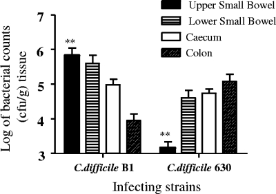
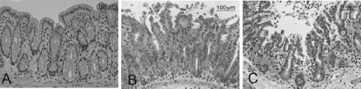
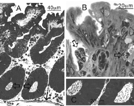
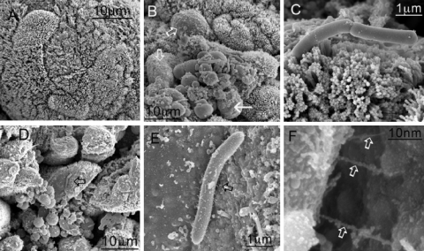
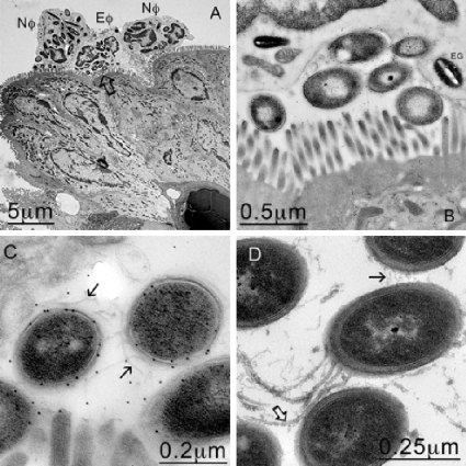
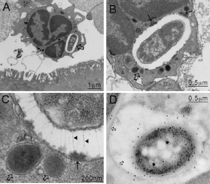
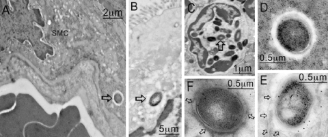
Currently, the Golden Syrian hamster is widely considered an important model of Clostridium difficile disease, as oral infection of this animal pretreated with antibiotics reproduces many of the symptoms observed in humans. Two C. difficile strains, B1 and 630, showed significant differences in the progression and severity of disease in this model. B1-infected hamsters exhibited more severe pathology and a shorter time to death than hamsters infected with 630. Histological changes in the gut did not correlate with absolute numbers of C. difficile bacteria, but there were clear differences in the distribution of bacteria within gut tissues. Light, scanning, and transmission electron microscopy revealed high numbers of B1 bacteria at the mucosal surface of the tissue, whereas 630 bacteria were more frequently associated with the crypt regions. Both B1 and 630 bacteria were frequently observed within polymorphonuclear leukocytes, although, interestingly, a space frequently separated B1 bacteria from the phagosome wall, a phenomenon not observed with 630. However, pilus-like structures were detected on 630 located in the crypts of the gut tissue. Furthermore, B1 bacteria, but not 630 bacteria, were found within nonphagocytic cells, including enterocytes and muscle cells.


Figures

FIG. 1.
FIG. 1.
Log bacterial counts recovered from different regions of the bowel following infection with approximately 100 spores of C. difficile B1 or 630. Tissue was obtained from the upper small bowel, the lower small bowel, the cecum, and the colon. The error bars indicate the standard deviations from the means for five animals. Asterisks indicate differences in colonization that are statistically significant.
FIG. 2.
FIG. 2.
Hematoxylin- and eosin-stained, paraffin-fixed sections of the upper colon of (A) uninfected hamsters, (B) 630-infected hamsters, and (C) B1-infected hamsters. In contrast to the results for the uninfected tissue, increasing numbers of polymorphs are present basally and at the surface of the mucosal of tissue infected with C. difficile 630, and the highest numbers are in tissue infected with C. difficile B1.
FIG. 3.
FIG. 3.
Semithin (1 μm) Lowicryl sections of tissue infected with C. difficile B1 and 630 and stained with toluidine blue. (A) In tissue infected with 630, bacteria are at the mucosal surface at the base of the crypts (open arrows) in close proximity to the basal lamina (solid arrow). (C) Higher magnification of bacteria in panel A. (B) In contrast, B1 bacteria were most often associated with PMNs (arrows).
FIG. 4.
FIG. 4.
SEM images of cecal tissue infected with C. difficile. Tissues were obtained 1 h after the onset of diarrhea (approximately 29 h postinfection for B1 and approximately 44 h postinfection for 630). (A) Control uninfected tissue. (B and D) Infection of the tissue with both 630 (B) and B1 (D) resulted in denuded patches of microvilli (open arrows), and organisms appeared to anchor themselves to the mucosal surface. (C and E) For 630, attachment was mediated largely at the pole of the organism (C), while for B1, the organisms appeared to be interacting with the surface by means of pilus-like structures between 200 and 300 nm long (E) (arrow). (F) Higher magnification of pilus-like structures in panel E (arrows).
FIG. 5.
FIG. 5.
PMNs engulfing C. difficile B1 at the mucosal membrane. (A) TEM image showing neutrophils (Nφ) and eosinophils (Eφ) at the mucosal surface that appear to have engulfed several bacteria. (B) Higher magnification of the area indicated by the arrow in panel A, confirming that C. difficile was trapped against the mucosal membrane by the host cells. (C) Higher magnification of panel B. The bacteria were confirmed to be C. difficile by immunogold labeling. (D) In contrast, 630 bacteria found in the crypts appear to express both flagella (open arrow) and pili (solid arrow).
FIG. 6.
FIG. 6.
(A) C. difficile B1 bacteria were most commonly found in association with PMNs within vacuoles (arrow). (B) Higher magnification of panel A. (C) When images of the vacuoles were enlarged (area indicated by open arrows in panels A and B), filamentous structures between 200 and 400 nm long were discerned (arrowheads). These structures appeared to pin back (solid arrows in panel B and solid arrow in panel C) the contents released from fusing lysosomes (open arrows in panels B and C), suggesting a possible mechanism for the cell to avoid direct contact with the digestive elements. (D) Immunogold labeling confirmed that the organisms inside the vacuole were C. difficile.
FIG. 7.
FIG. 7.
TEM images of tissue infected with C. difficile 630. Unlike B1 (Fig. 6), 630 seems to be unable to prevent killing within the neutrophils (A), and organisms located within phagosomes undergo degradation (B). In an enhanced image (D), the phagosomal membrane (open arrow) is located tightly around the bacteria, lysosomal contents are intimately associated with the bacteria, and a second dark vesicle is undergoing fusion (solid arrow) The bacteria within the phagosomal vesicles (C) and the flagellated cells within the crypt regions (E) were confirmed to be C. difficile by immunogold labeling with anti-630 antibody. In addition, antibody raised to PilA (CD3507) recognized the larger of the two structures expressed by free 630 bacteria located in the crypt regions, suggesting that the pili may be expressed in vivo (F).
FIG. 8.
FIG. 8.
C. difficile B1 was in several other locations within the gut, as determined by TEM. These locations included smooth muscle cells (A [arrow] and D), the mucosa (B [arrow] and E), and eosinophils (C [arrow] and F). In panels E and F the membrane around the bacteria is viable (arrows), but a membrane is not present in panel D (arrows).

References

    1. Barbut, F., B. Gariazzo, L. Bonne, V. Lalande, B. Burghoffer, R. Luiuz, and J.-C. Petit. 2007. Clinical features of Clostridium difficile—associated infections and molecular characterisation of strains: results of a restrospective study, 2000-2004. Infect. Control Hosp. Epidemiol. 28:131-139. - PubMed
    1. Bartlett, J. G., A. B. Onderdonk, R. L. Cisneros, and D. L. Kasper. 1977. Clindamycin associated colitis due to a toxin producing species of Clostridium in hamsters. J. Infect. Dis. 136:701-705. - PubMed
    1. Borriello, S. P., J. M. Ketley, T. M. Mitchell, F. E. Barclay, A. R. Welch, A. B. Price, and J. Stephen. 1987. Clostridium difficile—a spectrum of virulence and analysis of putative virulence determinants in the hamster model of antibiotic-associated colitis. J. Med. Microbiol. 24:53-64. - PubMed
    1. Calabi, E., F. Calabi, A. D. Phillips, and N. Fairweather. 2002. Binding of Clostridium difficile surface layer proteins to gastrointestinal tissues. Infect. Immun. 70:5770-5778. - PMC - PubMed
    1. Cerquetti, M., A. Serafino, A. Sebastianelli, and P. Mastrantonio. 2002. Binding of Clostridium difficile to Caco-2 epithelial cell line and extracellular matrix proteins. FEMS Immunol. Med. Microbiol. 32:211-218. - PubMed

Publication types

MeSH terms

LinkOut - more resources